Download
1 / 60
600 likes | 955 Vues
Classification of microorganisms and functional anatomy of prokatyotic and eukaroytic cells. CLASSIFICATION OF ORGANISMS. 1735 - Linnaeus-Established first classification system for classifying living things. Established the binomial*" system of nomenclature for_-naming organisms.

E N D
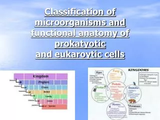
Classification of microorganisms and functional anatomy of prokatyoticand eukaroytic cells
CLASSIFICATION OF ORGANISMS • 1735 - Linnaeus-Established first classification system for classifying living things. • Established the binomial*" system of nomenclature for_-naming organisms. • This means that each living organism is given a * genus and species name. • This is called the scientific name and MUST always be written with the genus name first and the species name second.
The genus name must be capitalized and the species name written in lower case letters. • The entire scientific name must be either italicized or underlined.
Linaeus's classification system was composed of 2 Kingdoms • . A plant kingdom and an animal kingdom. • Microorganisms were considered so unimportant that they were grouped under a heading of Vermes in a catagory Chaos.
1969.Robert Whittaker-devised a five-kingdom classification system based on the 'cellular organization and nutritional patterns of organisms.
R. H. Whittaker • In 1969, R. H. Whittaker developed a 5 kingdom system which included: • Plants • Animals • Protist • Fungi • Monera/ Prokaryotes
ALL CELLS ARE CLASSIFIED AS EITHER PROKARYOTES OR EUKARYOTES • PROKARYOTES - pro means primitive • DNA contains no protein .lack nucleus • no organelles • single chromosome composed of DNA in loop. • chlorophyll, if present, is in cytoplasm. • ribosomes are smaller and free in the cytoplasm. • cell usually surrounded by a cell wall. • Reproduce by fission: no evidence of mitosis • EXAMPLES - bacteria, rickettsiae, chlamydiae, mycoplasms, cyanobacteria
EUKARYOTES- eu means true • have nucleus • have organelles (ex. mitochondria, lysosomes, Golgi apparatus,endoplasmic reticulum. • have multiple chromosomes in nucleus with protein around chromosome • ribosomes bound to membrane or free in cytoplasm. Larger. • chlorophyll, if present, is in organelle. • cell wall absent or less complex chemistry • reproduce by mitosis • EXAMPLE - fungi, protozoa, plants, animals
Cellular Components: • Plastids- contains pigment
NUCLEUS • double membrane boundcontains chromatin disappears during mitosis or meiosis when the chromosomes divide and is reformed after cytoplasmic organelle
NUCLEOLUS • Aka “little nucleus” • Found in the nucleus • Contains RNA and proteins for ribosome synthesis
CYTOPLASM • gelatin-like inside cell membrane • constantly flows • It contains the various organelles of the cell
ENDOPLASMIC RETICULUM • extensive convoluted membrane continuous with the outer nuclear membrane and enclosing a continuous internal space involved in the synthesis and transport of membrane proteins and lipids and of proteins destined for secretion from the cell
ENDOPLASMIC RETICULUM • A series of folded membranes that move materials (proteins) around in a cell like a conveyor belt • Smooth ER – ribosomes not attached to ER, functions in lipid synthesis • Rough ER – ribosomes attached to ER, functions in producing proteins
RIBOSOMES • Make proteins • Float freely or attached to the endoplasmic reticulum (ER) • Ribosomes are made in the nucleolus and are small particles of RNA
MITOCHONDRIA • Organelles that release energy from food (power house of cell) • This energy is released by AKA the powerhouse b/c they release energy(ATP) from food
Folds of mitochodria are called: cristae Mitochondria
GOLGI BODIES (GAWL jee) • Stacked flattened membranes • Sort and package proteins
LYSOSOMES (LI suh sohmz) • The word "lysosome" is Latin for "kill body." • The purpose of the lysosome is to digest things. They might be used to digest food or break down the cell when it dies. • Break down food molecules, cell wastes & worn out cell parts
VACUOLES • Temporary storage spaces • Store food, water, waste • may contain air, water, cell sap, partially digested food etc...
Centrioles • Short cylinder near nuclear envelope • There generally are 2 at right angles to each other • They control cell division
Peroxisome- • small organelle bounded by a single membrane and containing • catalase and peroxidases • important in detoxification reactions
Cell Wall • located beneath the capsule and external to the cytoplasmic membrane. All bacteria except for mycoplasms have cell walls. • Functions: • A. Maintain shape of bacteria • B. Prevent cell from rupturing - internal pressure 20 times externalpressure. • C. Point of anchorage for flagella
The cell wall is composed of peptidoglycan which is present either alone or in combination with other substances. • The peptidoglycan is composed of a carbohydrate backbone linked together by peptides.
Gram positive cell walls are composed of a thick layer of peptidoglycan approximately 25 nm in thickness. • Teiochoic acid is also present in the peptidoglycan layer. • The peptidoglycan makes up about 50% of the dry weight.
Gram negative cell walls are composed of a thin layer of peptidoglycan approximately 3 nm in thickness. • There is no teiochoic acid in the peptidoglycan layer but there are layers of lipopolysaccharide, phospholipids, and lipoproteins outside the peptidoglycan layer. • The peptidoglycan layer makes up about 10% of the dry weight.
Penicillin - antibiotic that functions in preventing the formation of cell walls. • Works best on Gram + bacteria. • Lysozyme - an enzyme found in body secretions that breaks down cell walls. Protoplast - a Gram + bacterium that has had the cell wall removed. • Made by treating cell with Lysozyme. • Spheroplast - a Gram - bacterium that has had the peptidoglycan layer of the cell wall removed. • Still has the cell membrane with the lipid layers of the cell wall on the outside. • Both protoplast and spheroplast will be spherical regardless of their original shape.
CELL MEMBRANE • approximately 7.5 nm thick and composed of a phospholipid bilayer with proteins dispersed throughout. • The phospholipid bilayer is composed of phosphates and glycerol on the outside with tails of fatty acids on the inside. • The polar heads are on the 2 outside surfaces and the non-polar tails are on the inside.
The arrangement of phospholipids and proteins is referred to as the fluid mosaic model due to the continuous change in the arrangement of the molecules. • Ethyl alcohol and poly myxin destroys the membrane.
Functions: • selective barrier - membrane is selectively permeable • serves as anchor for attachment of DNA • site of enzymes that function in cell wall synthesis (permeases) • site of enzymes that function in energy production • secretes exoenzymes that break down large molecules
MAJOR GROUPS OF MICROORGANISMS • BACTERIA – • unicellular, prokaryotic organisms that are among the most abundant on earth. • May be aerobic (requires oxygen) anaerobic (Needs no oxygen), or facultative (indifferent to oxygen, can survive either way). • Multiply by binary fission (single chromosome duplicates and 2 chromosomes move to different areas and membranes grow inward).
Bacteria have 3 basic shapes – • coccus-round • bacillus -rod • spiral-curved shape called spirillum • Some form endospores-cell shrinks, rounds up within old membrane and forms new thicker walls. • Average size is one micrometer. • BACTERIOLOGY-study of bacteria
Types of Bacteria • Rickettsiae • -small bacteria, among the tiniest microorganisms, usually transmitted by arthropods. Grow and multiply only within living cells. .5 urn. in size. Ex. Rocky Mountain Spotted Fever 1 micrometer = 1 thousandth of a millimeter • Chlamydiae • -subgroup of Rickettsiae, among smallest Rickettsiae, .25 urn. Grow and multiply only in living cells. Ex. disease of eye, lung, and urogenital tract. • Mycoplasms • smaller than Chlamydiae, .1 urn. Pleomorphic, has no cell wall, can grow on artificial media if certain steroids are added. Ex. walking pneumonia.
Types of Bacteria • Cyanobacteria • Known as blue-green algae. Nore closely related to bacteria because of structural and biochemical properties. • Not true bacteria. Possesses light-trapping pigments, many are blue but some are black, red, yellow, or green. Inhabit both fresh and salt water. Ex makes swimming pools green.
Protozoans: • Single celled eukaryotes that are usually microscopic. • Can cause parasitic diseases such as malaria. • Some are capable of photosynthesis but most are not. • They are classified according to their locomotion ( flagella, cilia, pseudopods) Protozoa average about 100um. • Protozoologyis the study of protozoas
FUNGI- • eukaryotic, non-photosynthetic, and contain chitin in their cell walls. Plants do not have chitin in the cell walls. Aid bacteria in decomposition. • Include yeast, molds, and mushrooms. Yeast are 8 urn and molds are 40 - 100 urn. • Mycology is the study of fungi.
4. ALGAE- • eukaryotic, photosynthetic plants that range in size from a single cell to giant kelp 30 m. in length. • One genus produces neurotoxins causing shellfish poisoning. • Microbiologist are interested in the algae classified in the kingdom Protista. • Extremely important in food chain in oceans. • Phycology is the study of algae.'
5. VIRUSES • -neither prokaryote or eukaryote. • Viruses are obligate intracellular parasites. • This means that the virus depends on the host'cell for reproduction and growth. • Viruses are non-cellular, infectious agents composed of a nucleic acid surrounded by a protein coat.
Viruses are considered the link between the living and the nonliving. • Cause diseases such as herpes, rabies, flu, hepatitis, polio, and chickenpox. • Only observable activity is reproduction. • Viruses are measured nanometers. Smallpox largest ( 250nm) polio smallest ( 20 nm) • Virology is the study of viruses.
Bacteria Morphology: the size, shape, structure and arrangement of cells. • SIZE - average one urn. • Because of small size have high surface area to volume ratio. This explains the high rate of growth and metabolism. Sphere has the smallest surface area to volume ratio. • SHAPE - three basic shapes governed by cell wall. • coccus - spherical, may be oval, elongated, or indented. • bacillus-rod • spiral-helically curved • a. vibrios-comma shaped • b. spirilia-rigid cell wall with flagella • c. spirochetes-flexible wall with no flagella
diplococcus - cells divide in one plane and remain attached in pairs. • streptococcus - cells divide in one plane and remain attached to form chains. • tetracoccus - cells divide in two planes and form groups of 4 cells. • staphylococcus - cells divide in three planes and produce grape like clusters of bacteria. • sarcinae - cells divide in three planes in a regular pattern producing cuboidal arrangement of cells. • Occasionally bacterial cells may be star shaped or square.
BACTERIAL SIRUCTURES Structures External to Cell Wall • 1. Glycocalyx-general term used for substances that surround the cell. • Viscous, gelatinous substance composed of polysaccharide, polypeptide, or both.