Download
1 / 67
690 likes | 1.06k Vues
Functional Anatomy of Prokaryotic and Eukaryotic Cells. Chapter 4. Comparison of Cell Types. Prokaryotic. Eukaryotic. DNA enclosed within a nucleus Multiple, linear molecules DNA associated with histone proteins Possess membrane-enclosed organelles Cell walls lack peptidoglycan

E N D
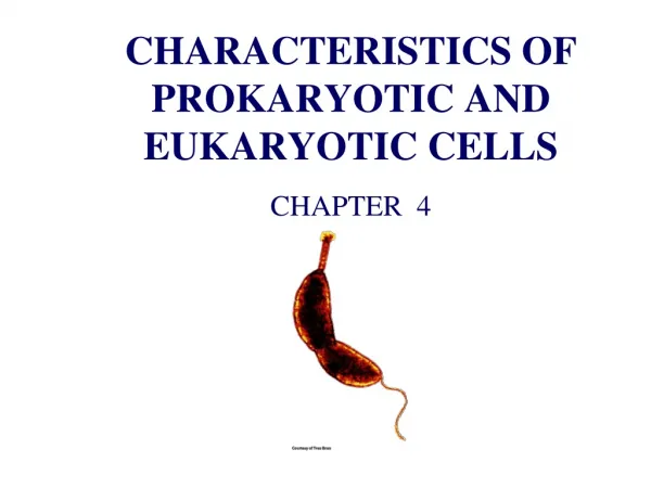
Functional Anatomy of Prokaryotic and Eukaryotic Cells Chapter 4
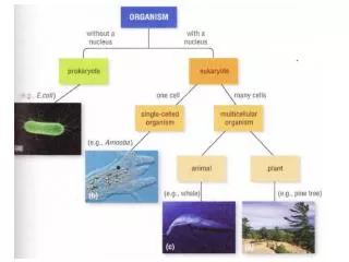
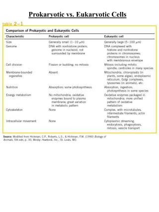
Comparison of Cell Types Prokaryotic Eukaryotic DNA enclosed within a nucleus Multiple, linear molecules DNA associated with histone proteins Possess membrane-enclosed organelles Cell walls lack peptidoglycan Divide by mitosis • DNA is not enclosed in a membrane • Often a single, circular molecule • DNA is not associated with histones • Lack membrane-enclosed organelles • Cell walls made of peptidoglycan • Divide by binary fission
Bacterial Shape is Hereditary • Most bacteria exhibit a single cell shape for their entire lives (Monomorphic) • Some exhibit a variety of shapes often dependent on the available nutrient in their environment • Pleomorphic
Cocci • Typically spherical cells • May remain attached after dividing • Diplococci divide in one plane • Streptococci form long chains • Tetrads divide in two planes • Sarcinae divide in three planes • Staphylococci form clusters resulting from non-uniform cell divisions
Rods (Bacilli) • Only divide along their short axis • May remain attached after dividing • Diplobacilli • Streptobacilli • Coccobacilli take on a rounded appearance as they age • Can be mistaken for cocci
Spirochetes • Vibrios are slightly curved • Spirilla are helical and fairly rigid in their structure • Spirochetes are also helical, but have fairly flexible structures • Move by means of axial filaments
Check Your Understanding • Distinguish between prokaryotic and eukaryotic cells. • Identify the three basic types of bacterial cell shapes. • Differentiate between the mechanism used by spirochetes and other bacteria for movement.
Glycocalyx Capsule Slime Layer Unorganized and loosely attached to the cell wall Composed primarily of polysaccharides Some polypeptides Helps keep bacteria hydrated in drier environs • Well-organized and firmly attached to the cell wall • Composed of polypeptides and polysaccharides • Important virulence factorby preventing phagocytosis • Bacillus anthracis • Streptococcus pneumoniae
Biofilms • Extracellular Polymeric Substance (EPS) formed by the glycocalyx of bacteria • Often results in a community of bacteria • Allows bacteria to adhere to surfaces in target environs and provides protection • Streptococcus mutans
Flagella • Long, filamentous appendages used to propel bacteria • H antigen is useful in identifying certain bacteria • Variety of arrangements: • Atrichous • Peritrichous • Monotrichous (Polar) • Lophotrichous (Polar) • Amphitrichous(Polar)
Prokaryotic Flagellar Structure • Filament: long, outer “tail” • Helical chains of flagellin • Hook: curved projection capable of rotation • Causes the filament to rotate in a clockwise or counterclockwise direction • Basal Body: anchors the hook and filament to the cell wall
Bacterial Movement via Flagella • Taxis is ability of a bacteria to move due to a stimuli • Chemotaxis: chemical • Phototaxis: light • Movement can be: • Positive – toward attractant • Negative – away from repellent • “Tumble” and “run”
Axial Filaments (Endoflagella) • Used by spirochetes for movement • Fibrils arise from ends of cell and spiral around the organism • Causes the microbe to spiral or “corkscrew” through the environment • Treponemapallidum(Syphillis) • Borreliaburgdorferi (Lyme disease)
Fimbriae • Composed of pilin • Often quite numerous • Aid bacteria in attaching to surfaces or other bacteria • Add to biofilm formation • Virulence factor • Neisseria gonorrheae • Escherichia coli O157:H7
Pili (Pilus) • Composed of pilin • Longer and much less numerous than fimbriae • Can be used to aid in gliding motility • Pseudomonas aeruginosaand Neisseria gonorrheae • Can be used to join cytoplasm of two bacteria permitting DNA exchange • Bacterial conjugation • Bacteria capable of forming a conjugation pilus are F+ • Can be used to transfer antibiotic resistance or other virulence factors
Check Your Understanding • Describe the structure and function of capsules and slime layers. • What is a biofilm and what is its importance in human medicine? • Differentiate between flagella, axial filaments, fimbriae, and pili. • Contrast the different types of flagellar arrangements used by bacteria for movement. • Describe taxis and differentiate between negative and positive taxis. Compare and contrast phototaxis and chemotaxis.
Peptidoglycan • Only present in bacteria • Adjacent sugars are linked polypeptides • “Peptido” + “glycan” • Protects bacteria from environmental stresses • Target for select antibiotics • Penicillin disrupts formation of peptide links • Leads to cell lysis
Gram (+) Cell Walls • Cell wall composed entirely of peptidoglycan • Forms a think wall around the cells • Additional compounds may be present • Teichoic acids • Aids in distinguishing certain groups of bacteria • Streptococcus sp.
Gram (-) Cell Walls • Thin layer of peptidoglycan forming the cell wall • Possess an outer membrane made of lipopolysaccharide • Prevents phagocytosis and action of complement • Resists action of antibiotics, digestive enzymes and bile • O antigen is useful in identifying certain bacteria
Atypical Cell Walls • A few bacteria lack a cell wall entirely • Possess sterols that help to stabilize their cell membranes • Some bacteria possess a high concentration of wax in their cell walls • Mycolic wax • Mycobacterium sp. Mycoplasma pneumoniae
Check Your Understanding • Compare and contrast cell walls of Gram-positive bacteria, Gram-negative bacteria, acid-fast bacteria, archaea, and mycoplasmas. • Why are drugs that target the peptidoglycan cell wall of bacteria useful? Why are mycoplasmas resistant to antibiotics that interfere with cell wall synthesis? • Compare and contrast archaea and mycoplasmas.
Plasma (Cell) Membrane • Fluid Mosaic Model • Phospholipid bilayer with integral and peripheral proteins • Proteins play a key role in transport, cell-cell binding, and enzymatic catalysis • Determines which materials may pass through the membrane • Selective permeability
Types of Transport Passive Active Movement of materials from areas of low to areas of high concentration Against the gradient Require use of ATP • Movement of materials from areas of high to areas of low concentration • With concentration gradient • No expenditure of ATP • Diffusion and Osmosis
Simple Diffusion • Passive transport of solute • Movement occurs until a dynamic equilibrium is reached • Movement of oxygen and carbon dioxide between cells
Facilitated Diffusion • Passive transport of solute • Requires use of membrane proteins • Channels for substances that cannot cross the membrane directly • Usually large or charged molecules • Sugars, vitamins • If still too large, bacteria secrete extracellualr enzymes to first digest the large particles
Osmosis • Passive transport of solvent • Water diffuses easily through membranes due to presence of aquaporins
Tonicity • Refers to the total amount of dissolved solute within an aqueous solution • Hypertonic solutions have more solutes (and less water) than cells • Isotonic solutions have the same amount of solutes as cells • Hypotonic solutions have less solutes (and more water) than cells
Check Your Understanding • Describe the structure, composition, and functions of the prokaryotic cell membrane. • Which chemical agents can cause injury to the bacterial cell membrane? • Differentiate between active and passive transport. • Differentiate between simple diffusion, facilitated diffusion, and osmosis. • Differentiate between hypertonic, isotonic, and hypotonic solutions. How does these affect cells?
Cytoplasm • ~80% water • Remainder is various organic molecules and ions • Thick, aqueous, semitransparent fluid • Provides the environment that all other structures are suspended within
Nucleoid • Portion of the cytoplasm where the bacteria’s genetic material is located • Contains a single, continuous, and (usually) circular double-stranded molecule of DNA • Additional, extra-chromosomal copies of DNA may also exist called plasmids • Often contain genes for virulence factors • Easily transferred between bacteria via conjugation
Ribosomes • Sites of protein synthesis • Each ribosome is actually made of two subunits • Each composed of rRNAand proteins • Large and small subunits must combine to form a functional ribosome • Target for select antibiotics • Stop protein synthesis in cell
Inclusions • Reserve deposits with various functions • Metachrome granules store phopshates for synthesis of ATP • Polysaccharide granules store glycogen and starch • Gas vacuoles help certain aquatic bacteria remain buoyant • Megnetosomes in certain bacteria act as magnets helping locate a surface for attachment
Endospores • Protective adaptation of certain Gram (+) bacteria to environmental stress • Bacillus sp. or Clastridium sp. • Bacteria alternate between a vegetative structure and a spore • Sporogenesis (sporulation) • Germination • Endospores are highly resistant to destruction
Check Your Understanding • Identify the functions of the nucleoid and ribosomes. • What are the general functions of inclusions? • Describe the functions of endospores, sporulation, and endospore germination. Under what conditions do endospores form?
Flagella and Cilia • Projections used for cell locomotion or movement of environment past the cell • Consist of microtubules • Formed by tubulin • 9 doublets surrounding 2 central microtubules (9 + 2) • Move in a wave-like pattern
Cell Wall and Glycocalyx • Most eukaryotic cells possess a cell wall • Cellulose in plants and some fungi • Chitin in fungi • Glucanand mannanin yeast • Protozoans possess a protein covering called a pellicle • Glycocalyx is found outside many animal cells • Extracellular Matrix • Proteins, sugars and lipids forming complex molecules • Anchor cells to surfaces and each other • Allows for cell-to-cell recognition
Plasma (Cell) Membrane • Phospholipid bilayer with various proteins and other stabilizing molecules interspersed • Similar in function to prokaryotic cell membranes • Differences include: • Types of proteins found within the membrane • Eukaryotic cells have carbohydrates used in cell recognition • Eukaryotic cells have sterols to strengthen the membrane • Phagocytosis is a process unique to eukaryotic cells