Download
1 / 47
480 likes | 789 Vues
The Respiratory System: Exchange of Gases. 0. 10. Respiration Takes Place Throughout the Body. Breathing (ventilation) : moving air in and out of lungs External respiration : gas exchange between air and blood in the lungs Internal respiration : gas exchange between blood and tissues

E N D
Respiration Takes Place Throughout the Body Breathing (ventilation): moving air in and out of lungs External respiration: gas exchange between air and blood in the lungs Internal respiration: gas exchange between blood and tissues Cellular respiration: oxygen use to produce ATP, carbon dioxide as waste
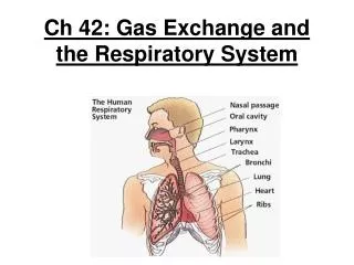
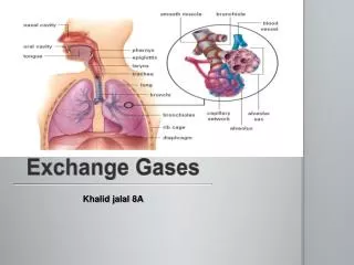
Respiratory System Consists of Upper and Lower Respiratory Tract Upper respiratory tract Nose, nasal passages, sinuses Pharynx Lower respiratory tract Larynx Trachea Bronchi and bronchioles Lungs Alveoli
Figure 10.1 Nasal cavity Filters, warms, and moistens air Nose Passageway for air UPPER RESPIRATORY TRACT Tongue Mouth Passageway for food and air Pharynx (Throat) Common passageway for air, food, and liquid Epiglottis Covers larynx during swallowing Larynx (Voice box) Production of sound Pleural membranes Cover the lungs and line the chest cavity Trachea (Windpipe) Main airway Lung Organ of gas exchange Bronchi Branching airways LOWER RESPIRATORY TRACT Alveoli Air sacs for gas exchange Intercostal muscle Moves ribs during respiration Right lung Left lung Rib Diaphram Skeletal muscle of respiration
Upper Respiratory Tract Filters, Warms, and Humidifies Air Functions of nose and pharynx Acts as passageway for respiration Has receptors for smell Filters larger foreign material from incoming air, inhaled microorganisms are entrapped in mucus Moistens and warms incoming air Has resonating chambers for voice
Figure 10.2 Sinuses Nasal cavity External nose Opening of the auditory tube Nostril Pharynx Tongue Epiglottis Glottis Larynx Trachea Esophagus
Lower Respiratory Tract Exchanges Gases Larynx Epiglottis: flexible flap of cartilage that routes air and food appropriately Vocal cords: assist in sound production Maintains an open airway Trachea Transports air to and from lungs Also known as the “windpipe” Kept open by C-shaped rings of cartilage Lined with mucus-secreting ciliated epithelium Traps foreign particles and moves them up and out of the lungs Cough reflex
Figure 10.3 Larynx Trachea Left bronchus Right bronchus Bronchioles Clusters of alveoli
Figure 10.4 Epiglottis Vocal cords Larynx Opening into larynx (glottis open) Closed glottis Upper trachea Position of the vocal cords during sound production. Position of the vocal cords during quiet breathing.
Figure 10.5 Epithelial tissue Connective tissue Cartilage rings Smooth muscle Relaxed state. The maximum diameter facilitates air movement in and out. During the cough reflex, the smooth muscle contracts briefly, reducing the diameter of the trachea. Combined with contraction of the abdominal muscles, this increases the velocity of air movement, forcibly expelling irritants or mucus from the trachea.
Lower Respiratory Tract Exchanges Gases Bronchi Trachea branches into two airways—right and left bronchi Contain ciliated epithelia, smooth muscle, cartilage Bronchioles Smaller branches, lack cartilage Functions of bronchi and bronchioles Air transport Clean, warm, and humidify incoming air Cleansing activity of cilia damaged by smoking leading to the development of smoker’s cough
Figure 10.6 Healthy airway. Smoker’s airway.
The Lungs Are Organs of Gas Exchange Supportive tissue enclosing the bronchi, bronchioles, blood vessels, and alveoli (air pockets where gas exchange occurs) Lungs are located in the thoracic cavity Each lung is enclosed in two layers of pleural membranes Area between pleural membranes (pleural cavity) contains fluid which reduces friction as lungs move Three lobes in right lung, two in left lung Lungs function relatively independently of each other
Figure 10.7 Pleural membrane lining thoracic cavity Rib Muscle Trachea Pleural cavity Pleural membrane attached to lung The three lobes of the right lung The two lobes of the left lung Diaphragm Heart (enclosed in pericardium)
Gas Exchange Occurs in Alveoli Alveoli: tiny air-filled sacs clustered at end of terminal bronchioles Walls of each alveolus are composed of only one squamous epithelial cell layer Combined surface area of alveoli: 800 ft2 Lipoprotein surfactant secreted by alveolar cells reduces surface tension enabling inflation of alveoli Premature infants may lack adequate surfactant and experience infant respiratory distress syndrome This may be successfully treated with surfactant therapy
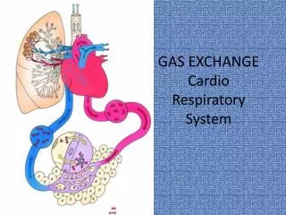
Pulmonary Capillaries Bring Blood and Air Into Close Contact Lungs receive deoxygenated blood from right ventricle of heart through pulmonary arteries Pulmonary capillaries allow blood to come into close proximity with air in alveoli Venules and veins collect the oxygenated blood from alveolar capillaries and return it to the left side of the heart for distribution throughout the body
Figure 10.8 Capillary Blood flow Air in aveolus O2 CO2 Bronchiole Small pulmonary vein Small pulmonary artery Bronchioles end in clusters of alveoli, each surrounded by capillaries. CO2 and O2 are exchanged across the capillary and alveolar walls by diffusion. Epithelial cell of alveolus Pulmonary venule Blood flow Blood flow Pulmonary arteriole Capillary network on surface of alveolus Photo of the surface of alveoli covered with capillaries.
Defenses of the Respiratory Tract Mucus—entraps microorganisms Cilia—push microorganisms and mucus up and out of respiratory tract Smoking—damages cilia, impairs this defense Smokers cough Cough reflex
The Process of Breathing Involves a Pressure Gradient Inspiration/expiration: air in/air out cycle 1. Relaxed state Diaphragm and intercostal muscles relaxed 2. Inspiration (inhale) Diaphragm contracts, pulling muscle down; intercostal muscles contract, elevating chest wall and expanding volume of chest, lowering pressure in lungs, pulling in air 3. Expiration (exhale) Muscles relax; diaphragm resumes dome shape; intercostal muscles allow chest to lower, resulting in increase of pressure in chest and expulsion of air
Figure 10.9 Air flows in No air movement Air flows out Ribs move upward and outward due to muscle contraction Ribs return to resting position External intercostals Lung volume increases, causing air pressure to fall Lung volume decreases, causing air pressure to rise Diaphragm Diaphragm contracts and flattens, moving downward Diaphragm relaxes 3 2 1 Expiration Inspiration Relaxed state
Lung Volumes and Vital Capacity Measure Lung Function Tidal volume Volume of air inhaled and exhaled in a single breath Dead space volume Volume of air that remains in the airways and does not participate in gas exchange Vital capacity Maximal volume that can be exhaled after maximal inhalation
Lung Volumes and Vital Capacity Measure Lung Function Inspiratory reserve volume Volume of air that can be inhaled beyond the tidal volume Expiratory reserve volume Volume of air that can be forcibly exhaled beyond the tidal volume Residual volume Volume of air remaining in the lungs, even after a forceful maximal expiration Measured by spirometer
Figure 10.10 A recording of lung capacity. After several normal breaths, the person inhales and then exhales maximally. The volumes indicated by the green line are for a normal person. The orange line is typical of a patient with emphysema. 6000 Normal person 5000 Person with emphysema Inspiratory reserve 4000 Vital capacity 3000 Lung volumes (ml) Tidal volume Expiratory reserve 2000 1000 Residual volume 0 Time Patient having his lung capacity determined with a spirometer.
Gas Exchange and Transport Occur Passively Partial pressure: the pressure exerted by one particular gas in a mixture of gases Partial pressure of a gas is proportional to its percentage of the total gas composition A gas always diffuses down its partial pressure gradient, from higher to lower partial pressure
External Respiration: The Exchange of Gases Between Air and Blood O2 diffuses from alveoli (PO2: 104 mmHg) into blood (PO2: 40 mmHg), down its partial pressure gradient CO2 diffuses from blood (PCO2: 46 mmHg) into alveoli (PCO2: 40 mmHg), down its partial pressure gradient
Internal Respiration: The Exchange of Gases with Tissue Fluids O2 diffuses down its pressure gradient from capillaries to interstitial fluid and then to cells CO2 diffuses down its pressure gradient from cells to interstitial fluid to capillaries The O2 and CO2 each flow down their own pressure gradient, in opposite directions
Figure 10.11 Slide 1 Dry inhaled air Moist exhaled air O2 160 O2 120 Breathing CO2 27 CO2 0.3 Alveolar air Alveolus 104 Pulmonary circulation 40 O2 104 O2 CO2 40 CO2 Lung capillaries 100 46 Capillary External respiration Pulmonary vein and aorta Transport O2 100 CO2 40 Systemic veins and pulmonary artery Internal respiration CO2 46 O2 40 100 O2 CO2 46 <40 >46 Systemic circulation Interstitial fluid surrounding cells Capillary networks in head, limbs, torso, and internal organs CO2 >46 O2 <40 Cells of tissues
Hemoglobin Transports Most Oxygen Molecules 98% of O2 in blood is carried bound to hemoglobin molecules in red blood cells Hb O2 HbO2 hemoglobin oxygen oxyhemoglobin 2% of O2 is dissolved in plasma
Most CO2 Is Transported in Plasma as Bicarbonate 70% of CO2 is converted to and transported in the plasma as bicarbonate 10% of CO2 is dissolved in plasma 20% of CO2 binds to hemoglobin for transport
Figure 10.12 Carbon dioxide transport in venous blood Oxygen transport in arterial blood Tissue cells 2% dissolves in plasma CO2 Alveous O2 CO2 20% combines with hemoglobin (Hb) CO2 O2 CO2 Blood is transported from lungs to tissue Hb O2 Hb 10% dissolves in plasma CO2 HbCO2 Enzyme H2O HbO2 Capillary H2CO3 Red blood cell 70% is converted to HCO3. HCO3 diffuses into plasma 98% combines with hemoglobin (Hb) H HCO3 Approximately 98% of all the O2 transported to the tissues by arterial blood is bound to hemoglobin (Hb) within red blood cells. Most of the CO2 transported to the lungs by venous blood is converted to bicarbonate (HCO3) within red blood cells and then is carried in plasma as dissolved HCO3. The rest is either dissolved in plasma as CO2 or transported within the red blood cells bound to hemoglobin.
Animation: The Human Respiratory System Right-click and select Play
The Nervous System Regulates Breathing Respiratory center is located in the medulla oblongata Establishes basic breathing pattern Neurons in medulla generate impulses every 4–5 seconds Stimulates intercostal muscles and diaphragm
Chemical Receptors Monitor CO2, H, and O2 Levels Monitoring of CO2 levels Receptors in medulla oblongata monitor CO2 levels indirectly Receptors monitor H ions in cerebrospinal fluid A rise in PCO2 in arterial blood will cause a rise in H in cerebrospinal fluid, which will signal medulla oblongata to increase respiratory rate This is the primary regulator of respiration under normal conditions
Chemical Receptors Monitor CO2, H, and O2 Levels Monitoring PO2 Aortic and carotid bodies monitor PO2 Become activated if PO2 falls by at least 20% Will increase rate and depth of breathing in response to sufficiently lowered arterial PO2 The rate and depth of normal breathing is determined by the need to get rid of CO2 rather than the need to obtain O2
We Can Exert Some Conscious Control Conscious control resides in the cerebral cortex Able to modify breath to speak and sing Able to hold breath temporarily, but can’t override automatic controls located in medulla oblongata indefinitely
Figure 10.13 The respiratory center: establishes a regular pattern of breathing 3 Higher centers in the cortex can exert conscious control over respiration. 1 Receptor cells near the respiratory center respond to changes in cerebrospinal fluid H caused by increases in arterial CO2. Medulla oblongata 2 Receptor cells in the carotid and aortic bodies respond to large decreases in arterial O2. Carotid bodies Aortic bodies Intercostal muscles Diaphragm
Disorders of Respiratory System Reduced air flow or gas exchange Asthma: spasmodic contraction of bronchi Caused by episodic allergic responses Controlled by bronchodilators and corticosteroids Emphysema: alveoli permanently impaired Breathlessness due to decreased surface area for gas exchange Most cases result from smoking
Disorders of Respiratory System Reduced air flow or gas exchange (cont’d) Bronchitis: inflammation of bronchi May be acute or chronic Most cases related to smoking or air pollution Cystic Fibrosis: an inherited condition Abnormally thick mucus in lungs impedes air flow Frequent pulmonary infections Other organ systems may be affected
Microorganisms Can Cause Respiratory Disorders Upper respiratory infections Colds and flu Caused by viruses Antibiotics not effective Pneumonia: infection of the lungs May be caused by bacteria or viruses One of the top 10 causes of death in the United States Treatment depends on cause
Microorganisms Can Cause Respiratory Disorders Respiratory infections and intoxications Tuberculosis: bacterial infection that scars the lungs May be active infection with symptoms, or dormant infection that will reactivate later Diagnosis: tuberculin skin test and chest X-ray Treatment: antibiotics Botulism: poisoning by bacterial toxin Toxin consumed in improperly preserved foods Causes paralysis of skeletal muscles including intercostals and diaphragm
Lung Cancer Is Caused By Proliferation of Abnormal Cells Cancer: uncontrolled growth of abnormal cells Impairs air flow, gas exchange, blood flow 1/3 of all U.S. cancer deaths Causes 90% of cases associated with smoking Radon gas Workplace chemicals such as asbestos
Figure 10.14 Cancerous lung. Normal lung.
Exposure to Asbestos Can Lead to Mesothelioma Mesothelioma Deadly cancer of lining of lungs, heart, and abdomen Most cases associated with asbestos exposure Symptoms appear 20–50 years after exposure Often undiagnosed until well-established Life expectancy after diagnosis: 8–14 months
Pneumothorax and Atalectasis: A Failure of Gas Exchange Pneumothorax: collapse of one or more lobes of the lungs Most often due to penetrating wound Inability to inflate the lung results in poor gas exchange Treatment: remove air from pleural cavity Atalectasis: lack of gas exchange due to alveolar collapse or fluid build-up Can be a complication of surgery May result from deficient surfactant
Congestive Heart Failure Impairs Lung Function Congestive heart failure: heart becomes a less efficient pump Failure of left side of heart Blood pressure increases within pulmonary capillaries Fluid build-up occurs in pulmonary interstitial spaces and alveoli Impaired gas exchange Treatment: improve pumping action of heart and remove fluid