Download
1 / 32
420 likes | 943 Vues
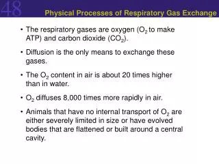
GAS EXCHANGE Cardio Respiratory System. GAS EXCHANGE. supplies oxygen for aerobic cellular respiration (reactant) 2. removes carbon dioxide from aerobic cellular respiration (product) 3 . M ust carry out ventilation - actively moving air in and out of body surfaces

E N D
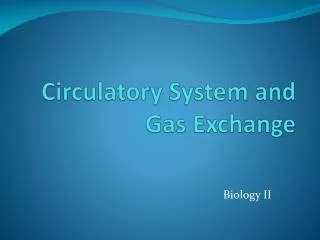
GAS EXCHANGE • supplies oxygen for aerobic cellular respiration (reactant) 2. removes carbon dioxide from aerobic cellular respiration (product) 3.Must carry out ventilation - actively moving air in and out of body surfaces 4. Terrestrial – gases in air Aquatic – gases dissolved in water
SOME AQUATIC INVERTEBRATES: Thin-wall - Gases diffuse through the membrane Circular canal Fig. 42-2 Mouth Pharynx Mouth Radial canal 5 cm 2 mm (a) The moon jelly Aurelia, a cnidarian (b) The planarian Dugesia, a flatworm
OTHER AQUATIC INVERTIEBRATES USE GILLS Fig. 42-21 Coelom Gills Gills Tube foot Parapodium (functions as gill) (a) Marine worm (c) Sea star (b) Crayfish
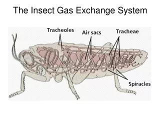
Air sacs Tracheae Fig. 42-23 External opening Tracheoles Mitochondria Muscle fiber Body cell Air sac Tracheole Trachea Body wall Air 2.5 µm
GILLS Fluid flow through gill filament Oxygen-poor blood Anatomy of gills Oxygen-rich blood Gill arch Lamella Fig. 42-22 Gill arch Gill filament organization Blood vessels Water flow Operculum Water flow between lamellae Blood flow through capillaries in lamella Countercurrent exchange PO2 (mm Hg) in water 150 120 90 60 30 Gill filaments Net diffu- sion of O2 from water to blood 110 80 50 20 140 PO2 (mm Hg) in blood
LUNGS Air Air Anterior air sacs Fig. 42-26 Trachea Posterior air sacs Lungs Lungs Air tubes (parabronchi) in lung 1 mm EXHALATION Air sacs empty; lungs fill INHALATION Air sacs fill
Rib cage expands as rib muscles contract Rib cage gets smaller as rib muscles relax Air inhaled Air exhaled Fig. 42-25 Lung Diaphragm INHALATION Diaphragm contracts (moves down) EXHALATION Diaphragm relaxes (moves up)
Cerebrospinal fluid Pons Fig. 42-27 Breathing control centers Medulla oblongata Carotid arteries Aorta Diaphragm Rib muscles
Inhaled air Exhaled air Alveolar spaces Alveolar epithelial cells CO2 O2 CO2 O2 Alveolar capillaries of lung Pulmonary veins Pulmonary arteries Fig. 42-UN2 Systemic veins Systemic arteries Heart Systemic capillaries O2 CO2 O2 CO2 Body tissue
Alveolus Alveolus PCO2 = 40 mm Hg PO2 = 100 mm Hg Fig. 42-28 PO2 = 40 PCO2 = 46 PCO2 = 40 PO2 = 100 Circulatory system Circulatory system PO2 = 40 PO2 = 100 PCO2 = 40 PCO2 = 46 PO2 ≤ 40 mm Hg PCO2 ≥ 46 mm Hg Body tissue Body tissue (a) Oxygen (b) Carbon dioxide
Chains Fig. 42-UN1 Iron Heme Chains Hemoglobin
100 O2 unloaded to tissues at rest 80 O2 unloaded to tissues during exercise 60 Fig. 42-29a O2 saturation of hemoglobin (%) 40 20 0 20 40 80 100 0 60 Tissues during exercise Tissues at rest Lungs PO2 (mm Hg) (a) PO2 and hemoglobin dissociation at pH 7.4
100 pH 7.4 80 pH 7.2 Fig. 42-29b Hemoglobin retains less O2 at lower pH (higher CO2 concentration) 60 O2 saturation of hemoglobin (%) 40 20 0 0 20 40 60 80 100 PO2 (mm Hg) (b) pH and hemoglobin dissociation
100 Fetus 80 Fig. 42-UN3 Mother O2 saturation of hemoglobin (%) 60 40 20 0 100 0 20 40 60 80 PO2 (mm Hg)
Body tissue CO2 transport from tissues CO2 produced Interstitial fluid CO2 Fig. 42-30a Capillary wall CO2 Plasma within capillary CO2 H2O Hemoglobin picks up CO2 and H+ Red blood cell H2CO3 Hb Carbonic acid HCO3– Bicarbonate H+ + HCO3– To lungs
CO2 transport to lungs HCO3– HCO3– H+ + Hemoglobin releases CO2 and H+ Fig. 42-30b Hb H2CO3 H2O CO2 Plasma within lung capillary CO2 CO2 CO2 Alveolar space in lung