Download
1 / 33
390 likes | 866 Vues
Physical Processes of Respiratory Gas Exchange. The respiratory gases are oxygen (O 2 to make ATP) and carbon dioxide (CO 2 ). Diffusion is the only means to exchange these gases. The O 2 content in air is about 20 times higher than in water. O 2 diffuses 8,000 times more rapidly in air.

E N D
Physical Processes of Respiratory Gas Exchange • The respiratory gases are oxygen (O2 to make ATP) and carbon dioxide (CO2). • Diffusion is the only means to exchange these gases. • The O2 content in air is about 20 times higher than in water. • O2 diffuses 8,000 times more rapidly in air. • Animals that have no internal transport of O2 are either severely limited in size or have evolved bodies that are flattened or built around a central cavity.
Physical Processes of Respiratory Gas Exchange • Fick’s law of diffusion: Q = DA (P1 - P2/L) • Q is the rate at which a substance diffuses between two locations. • D is the diffusion coefficient. • A is the cross-sectional area over which the substance is diffusing. • P1 and P2 are the partial pressures of the gas at two locations. • L is the distance between these locations. • Diffusion depends on Partial pressure (p) of the gases, Area, and Diffusion length • In Atmosphere - pOxygen (21%)> pCarbon dioxide (.03%)
Physical Processes of Respiratory Gas Exchange • Animals maximize the diffusion coefficient by using air rather than water for diffusion whenever possible. • Other adaptations for maximizing respiratory gas exchange must influence the surface area for exchange (A) or the partial pressure gradient across that surface area [(P1 – P2)/L].
Figure 48.3 Gas Exchange Systems Anatomical adaptations to maximize the surface area for gas diffusion (A in Fick’s law) include external and internal gills and lungs
Adaptations for Respiratory Gas Exchange • Driving diffusion of gases across gas exchange membranes (i.e., maximizing the partial pressure gradients—(P1 – P2)/L in Fick’s law) is accomplished in several ways: • Thin membranes shorten the diffusion path (L). • Ventilation brings in fresh air with the high PO2 and the low PCO2. • Perfusion by the circulatory system helps maintain the low PO2 and the high PCO2 on the inside of exchange surfaces.
Adaptations for Respiratory Gas Exchange • The perfusing blood flow on the inner surface of the lamellae is unidirectional. • Afferent (to gills) and efferent (away from gills) blood vessels ensure a countercurrent flow to maximize the PO2 gradient.
Figure 48.6 Countercurrent Exchange Is More Efficient than Concurrent Exchange
Figure 48.7 The Respiratory System of a Bird (Part 1) Air flows unidirectionaly
Figure 48.8 The Path of Air Flow through Bird Lungs (Part 1)
Figure 48.8 The Path of Air Flow through Bird Lungs (Part 2)
Adaptations for Respiratory Gas Exchange • In mammal lungs, ventilation is tidal: Air flows in and out by the same route. • At rest, the amount of air exchanged is the tidal volume. • The additional volume of air taken in by inhaling deeply is the inspiratory reserve volume. • The additional volume we can exhale is the expiratory reserve volume. • The total of these three volumes in the vital capacity.
Gas Exchange in Human Lungs • The Bronchioles end in the alveoli which are thin-walled air sacs and are the sites of gas exchange. • Capillary blood vessels closely surround the alveoli, resulting in a diffusion path of less than 2 mm, which is less than the diameter of a red blood cell.
Gas Exchange in Human Lungs • Two adaptations that aid the breathing process in mammals are mucus and surfactants. • Cells lining the airways produce a sticky mucus that captures dirt and microbes. • This mucus is cleared by cilia beating upward toward the trachea and pharynx, where it is swallowed.
Gas Exchange in Human Lungs • A surfactant is a chemical substance that reduces the surface tension of a liquid. • The aqueous lining of the lung has surface tension that must be overcome to permit inflation. • Cells in the alveoli produce surfactant molecules when they are stretched. • Premature babies may develop respiratory stress syndrome if they are born before cells in the alveoli are producing surfactant.
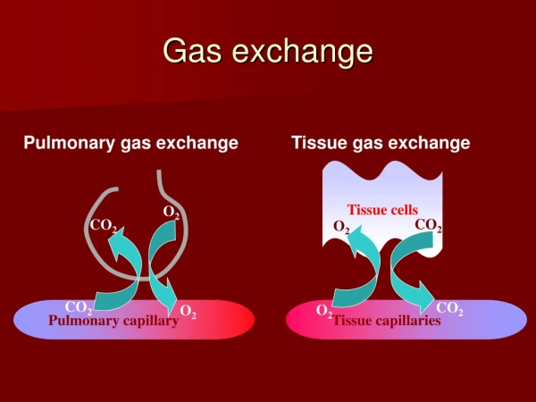
Blood Transport of Respiratory Gases • Ventilation and perfusion work together. Ventilation delivers O2 to the environmental side of the exchange surface; perfusion delivers CO2 to the exchange surface, where it diffuses out and is swept away by ventilation. • As O2 diffuses from the alveoli into the blood, it is swept away and delivered to the cells and tissues of the body. • Most O2 is carried by the oxygen-binding pigment, hemoglobin, in red blood cells. • Hemoglobin has 60 times the capacity of plasma to transport O2.
Blood Transport of Respiratory Gases • The influence of pH on the function of hemoglobin is known as the Bohr effect. • This effect occurs when the pH of the blood falls and the H+ ions bind to hemoglobin and decrease its affinity for O2. • The oxygen-binding curve shifts to the right. • The hemoglobin will then release more O2 to the tissues where pH is low.
Blood Transport of Respiratory Gases • Another regulator of hemoglobin function is 2,3 bisphosphoglyceric acid (BPG). • In red blood cells BPG combines with deoxygenated hemoglobin and causes it to have a lower affinity for O2. • The result is that the hemoglobin releases more of its bound O2 to tissues than usual. • If a person goes to a high altitude or starts exercising, the level of BPG goes up, and hemoglobin releases more O2 where it is needed.
Regulation of Breathing • Breathing is controlled by the autonomic nervous system. • The brain stem generates and controls the breathing rhythm. • Groups of neurons within the medulla increase their firing rate just prior to inhalation. • With increased firing, the diaphragm contracts and inhalation occurs. • When the firing stops, the diaphragm relaxes, and exhalation occurs. • Exhalation is actually a passive elastic recoil of lung tissue. When breathing demands are high, as during exercise, the motor neurons for the intercostal muscles are fired to increase inhalation and exhalation volumes. • Brain areas above the medulla (Pons) modify breathing to allow speech, eating, coughing, and emotional states.
Regulation of Breathing • CO2 sensors (monitor pH high CO2-Low pH) are located on the medulla surface near the neurons that generate the breathing rhythm. • However O2 sensors are also in tissue nodes on the aorta and carotid arteries called carotid and aortic bodies. • If PO2 of blood drops, or if blood pressure drops, chemoreceptors in the bodies send nerve impulses to the brain breathing center.