Download
1 / 53
530 likes | 605 Vues
Explore how DNA is condensed and packaged in chromosomes, the structure of human chromosomes, histones, nucleosomes, and higher-order compaction in eukaryotic cells. Learn the essential concepts of genetics and DNA organization.

E N D
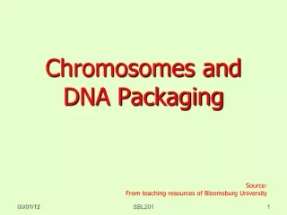
Chromosomes and DNA packaging Dr. Maryam Tahmasebi Birgani PhD in molecular genetics Tarbiat Modares University of Tehran University of Florence, Italy
Genetic Material in the Living Cells • Cells contain a nucleus surrounded by a nuclear membrane in eukaryotic cells, and a nuclear region in the prokaryotic cells. • In a non-dividing cell the nucleus is filled with a thread-like material known as "chromatin". • Chromatin is made up of DNA and proteins (mainly histones and some non-histone acidic proteins).
The Normal Human Chromosomes • Normal human cells contain 23 pairs of homologous chromosomes: • 22 pairs of autosomes. • 1 pair of sex chromosomes. • Autosomes are the same in males and females • Sex chromosomes are: • XX in females • XY in males. • Both X are homologous. Y is much smaller than X and has only a few genes.
Chromosome • One member of each chromosome pair is derived from each parent. • Somatic cellshave diploid complement of chromosomes i.e. 46. • Germcells(Gametes: sperm and ova) have haploidcomplement i.e 23. • Individual chromsomes are recognized by • arm lengths ( p- short, q -long). • centromere position (metacentric, sub-metacentric, acrocentric, telocentric).
DNA Condensation: Why?A severe problem of packaging • Largest human chromosome: ~3 x 10⁸ bp • A typical cell = 10 μm= 10 x 10-6 m • Therefore the DNA must be compacted ~104-fold.
Histone octamersOrganize DNA into Repeating Units • The first evidence that DNA is packaged into regularly organized units came from studies in which chromosomal DNA was treated with a nonspecific DNA nuclease, that cuts DNA wherever it is not associated with proteins. • The digested DNA fragments were then analyzed for size in an agarose gel. • If DNA is packaged by proteins into units of a particular size, the nuclease would cleave the DNA between these units, and the protected DNA segments would migrate in the gel as a ladder of unit-sized bands. • The results of such experiments revealed a series of regularly spaced DNA bands about 200 bp apart, indicating that DNA is packaged by proteins into units that encompass approximately 200 bp
The earliest evidence of DNA packaging. Isolated chromatin was treated with nuclease and analyzed by agarose gel electrophoresis. The result was a DNA ladder of fragments that differed in length by 200 bp, suggesting that DNA packaging involves a repeat unit of 200 bp. [Source: Roger Kornberg, MRC Laboratory of Molecular Biology.]
… • Consist of 5 classes: H1, H2A, H2B, H3, H4. • When the protein-DNA units (nucleosomes) were examined by SDS−polyacrylamide gel electrophoresis (SDS-PAGE), four histone proteins (H2A, H2B, H3, and H4) were found in approximately equimolar ratios . • A fifth histone (H1) was present in about half the amount relative to the other four histones. H1 is Lys rich. • The five histones have molecular weights (Mr) between 11,000 and 21,000.
Histones are rich in the basic amino acids arginine and lysine, which together make up about 25% of the amino acid residues in any given histone protein. • Histone proteins are highly conserved among eukaryotic cells. • Histones H3 and H4 are nearly identical in all eukaryotes, suggesting strict conservation of their functions. • Histones H1, H2A, and H2B show less sequence similarity, but on the whole, they are more conserved than other types of proteins. • Salt bridges between positively charged histones and negatively charges DNA play a major role in stabilizing DNA-histone complex
Nucleosome (10 nm diameter): 8 histones in bead & 1 outside. Each bead: is surrounded by 140 bpDNA and there are 60 bp in the linker region. Space between beads is about 14 nm.
… • Higher-order DNA compaction in a eukaryotic chromosome. This model shows the levels of organization that could provide the observed degree of DNA compaction in the chromosomes of eukaryotes. • The DNA is wrapped around histone octamers • H1 stimulates formation of the 30 nm filament. • Further levels of organization are not well understood but seem to involve further coiling and loops in the form of rosettes, which also coil into thicker structures. • progressive levels of organization take the form of coils upon coils upon coils. It should be noted that in cells, the higher-order structures (above the 30 nm filament) are unlikely to be as uniform as depicted here.
Eukaryotes contain thousands of times more DNA than do bacteria, and as a result, the DNA–condensation problems of eukaryotes—compacting the DNA so that it fits in the cell nucleus—are more complex than those of bacteria. • Bacteria do not contain nucleosomes, although they have small, basic (positively charged) proteins that are involved in condensing their DNA.
DNA compaction must be dynamic, because changes in the degree of condensation must occur quickly and when needed, as the cell passes through the stages of the cell cycle. • Furthermore, when in its most highly compacted form, DNA is not accessible to transcription or replication enzymes, so it must be able to rapidly expose regions containing genes that are required at any given moment, and then condense again. • Modification enzymes that alter the state of DNA condensation, and can target their activity to specific regions of the chromosome that must be transcribed or replicated.
Chromatin is divided into euchromatin and heterochromatin • Individual chromosomes can be seen only during mitosis. • During interphase, the general mass of chromatin is in the form of euchromatin. • Euchromatin is less tightly packed than mitotic chromosomes. • Regions of heterochromatin remain densely packed throughout interphase.
Essential for being as a chromosome • Centromere (Repeated DNA) • Movement of chromosomes during cell division • Telomere ( TTAGGG repeats) • Structure of chromosome • Control of cell division • Cell senescence or aging
Chromosomes have banding patterns • Certain staining techniques cause the chromosomes to have the appearance of a series of striations called G-bands. • The bands are lower in G • C content than the interbands. • Genes are concentrated in the G • C-rich interbands.
Chromosome ideogram: Arm (p and q) Region ( p:2 and q:3) Band Sub-Band “ Chromosome No. + Arm+ Region+ Band+ Sub-band “ Example: 15q11-q16
Chromosome Banding • Chromosome banding methods are either based on staining chromosomes with a dye or on assaying for a particular function. • The most common methods of dye based chromosome banding are • Bands that show strong staining are referred to as positive bands; weakly staining bands are negative bands. • G-positive bands are usually just called G-bands and likewise for R positive (R-) bands. Positive C-bands contain constitutive heterochromatin. Q-bands are considered equivalent to G-bands.
The metaphase chromosomes are treated with trypsin (to partially digest the chromosome) and stained with Giemsa stain. • Heterochromatic regions, which tend to be rich with adenine and thymine (AT-rich) DNA and relatively gene-poor, stain more darkly in G-banding. • In contrast, less condensed chromatin—which tends to be rich with guanine and cytosine (GC-rich) and more transcriptionally active—incorporates less Giemsa stain, and these regions appear as light bands in G-banding. • The pattern of bands are numbered on each arm of the chromosome from the centromere to the telomere
Chromosome Abnormality Aberration Aneuploidy Hetroploidy Numerical Chromosomal Euploidy Abnormality Deletion ;del Iso chromosome ; iso, i Duplication; dup Structural Inversion ; inv Ring chromosome; r Translocation ; t
Numerical chromosome aberration • Aneuploidy An aneuploid is an individual organism whose chromosome number differs from the wild type by part of a chromosome set. Generally, the aneuploid chromosome set differs from wild type by only one or a small number of chromosomes. Aneuploids can have a chromosome number either greater or smaller than that of the wild type: Trisomics (2n + 1) Double Trisomy (2n+1+1) Tetrasomics (2n+2) Monosomics (2n − 1) Nullisomics (2n − 2) Double Monosomics (2n-1-1)
Trisomy • 2n+1 • Down syndrome : Trisomy 21 • Patau syndrome: Trisomy 13 • Edwards syndrome: Trisomy 18 • Trisomy 16 : common reason of miscarriage in first trimester • Chromosome Non-disjunction (Meiosis)
Chromosome Non-disjunction n n+1 n+1 n
Monosomy • 2n-1 • Lethal phenotype • Non-disjunction • Anaphase lag • Turner syndrome
Translocation • In genetics, a chromosome translocation(t) is a chromosome abnormality caused by rearrangement of parts between non-homologous chromosomes. • Reciprocal translocation (rcp): • Reciprocal translocations are usually an exchange of material between non-homologous chromosomes. • Estimates of incidence range from about 1 in 500 to 1 in 625 human newborns.
classification In a balanced translocation, pieces of chromosomes are rearranged but no genetic material is gained or lost in the cell. An unbalanced translocation occurs when a child inherits a chromosome with extra or missing genetic material from a parent with a balanced translocation.
Robertsonian (rob): • This is a type of nonreciprocal translocation caused by breaks at or near the centromeres of two acrocentric chromosomes. • acrocentric chromosomes: chromosome 13, 14, 15, 21 and 22. • Centric Fusion • The reciprocal exchange of parts gives rise to one large metacentric chromosome and one extremely small chromosome that may be lost from the organism with little effect because it contains so few genes. • The resulting karyotypein humans leaves only 45 chromosomes, since two chromosomes have fused together.
Deletion • A part of a chromosomeor a sequence of DNA is missed. • Deletion symbol: del • Classification: • Terminal • Interstitial • Cri du chat syndrome:5p- • Angel man syndrome: 15q
Cri-du-chat (cat's cry) syndrome, also known as 5p- (5p minus) syndrome, is a chromosomal condition that results when a piece of chromosome 5 is missing. • Infants with this condition often have a high-pitched cry that sounds like that of a cat. • intellectual disability • delayed development, • small head size (microcephaly) • low birth weight • weak muscle tone (hypotonia) in infancy • Affected individuals also have distinctive facial features, including widely set eyes (hypertelorism) • low-set ears, • a small jaw • a rounded face. • Some children with cri-du-chat syndrome are born with a heart defect.
A child with Angelman syndrome will begin to show: • signs of delayed development at around 6-12 months • they may not speak at all or may only be able to say a few words. • They may have difficulty walking because of problems with balance and co-ordination (ataxia). • Their arms may tremble or move jerkily, and their legs may be stiffer than normal. • A number of distinctive behaviours are associated with Angelman syndrome. These include: • frequent laughter and smiling, often with little stimulus • being easily excitable, often flapping the hands • being restless (hyperactive), • having a short attention span • problems sleeping and needing less sleep than other children
Inversion Centromere is not involved Centromere is involved
Paracentric Inversion Pericentric Inversion