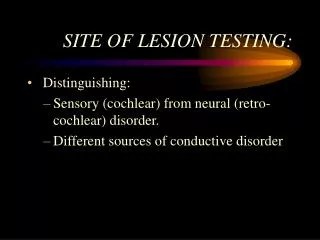
SITE OF LESION TESTING:
380 likes | 1.05k Vues
SITE OF LESION TESTING:. Distinguishing: Sensory (cochlear) from neural (retro-cochlear) disorder. Different sources of conductive disorder. MEASURES OF SUCCESS: SENSITIVITY. Percentage of persons with a disorder who show up on your test as having that disorder.

SITE OF LESION TESTING:
E N D
Presentation Transcript
SITE OF LESION TESTING: • Distinguishing: • Sensory (cochlear) from neural (retro-cochlear) disorder. • Different sources of conductive disorder
MEASURES OF SUCCESS: SENSITIVITY • Percentage of persons with a disorder who show up on your test as having that disorder. • In this application, % of persons with neural disorder that show a “neural result” on the site of lesion test.
MEASURES OF SUCCESS: SPECIFICITY • percentage of persons without a disorder who show up on your test as not having that disorder. • In this application, % of persons with a cochlear disorder (or no auditory disorder at all) who show up on your test as not having any neural disorder.
Loudness Recruitment Tests • Based on the changes in loudness perception that accompany different auditory disorders.
Recruitment: • "Abnormal growth of loudness" or, persistence of normal loudness above threshold. More common at higher frequencies.
Recruitment is consistent with cochlear damage • from noise • ototoxic substances • aging • and other causes
Decruitment: • Abnormal impairment of loudness growth • loudness curve actually moves away from normal line • lack of functioning nerve cells to code intensity • associated with retro-cochlear (VIIIth n.) lesions.
The Alternate Binaural Loudness Balance(ABLB)Test • requires: • - normal hrg in one ear at freq to be used • - difference in between ears > 25 dB
ABLB • tones pulse alternating between ears 2 or 3 times per judgement. • pt is asked which ear is louder or same • - begin at 20 SL in poorer ear, • - 0 SL in better ear. • - adjust level in better ear 5 dB steps.
ABLB • - find level where loudness judged equal. • - increase poorer ear by 10 or 20 dB and repeat adjustments in better ear.
PLOTTING ABLB RESULTS: • Use the “LADDERGRAM” • Connect decibel values judged equally loud
ABLB SUCCESS? • Sensitivity = 51% • Specificity = 88%
The Alternate Monaural LB (AMLB) Test • tone alternates between 2 frequencies in the same ear. • judgment and procedure is similar to ABLB, • but comparing "the high pitch versus the low pitch.” • generally this is harder for people to do.
Differential Intensity Discrimination • The Short Increment Sensitivity Index (SISI) • The High Level SISI
TheShort Increment Sensitivity Index • detection of brief (200 ms) 1 dB-increments in a 20 SL tone • 20 trials • > 70 % = cochlear damage • < 30 % = other damage or normal
B. High Level SISI • at 75 dB HL • Results: • > 70 % = normal or cochlear • < 30 % = retrocochlear
SISI SUCCESS? • Sensitivity = 68% • Specificity = 90%
Tone Decay: • Loss of audibility for a tone that is on continuously. • Greater decay is indicative of retrocochlear problem. • There are different methods:
Some Tone Decay Tests • Carhart: begin at 0 SL, up in 5 dB steps until tone is heard for a full minute • Olson-Noffsinger: begin at 20 SL, up until heard for full minute.
Tone Decay Results: • Type I: no decay: norm, conduct or cochlear • Type II: heard for longer times as level is increased: cochlear • Type III: No growth with increasing level: retrocochlear
TONE DECAY SUCCESS? • Sensitivity = 75% • Specificity = 91%
Bekesy Audiometry: • Pt. controls level of tone, • Continuous tone: tone on constantly (C) • Interrupted tone: pulsed on and off (I) • Adaptation should only occur for C, not I
Bekesy Results: I: C and I overlap: norm or cond. II: C below I at freqs of HL: Cochlear III: I follows loss, C drops to bottom: Retro IV: C below I by 20-25 dB: Coch or Ret V: I below C: False hearing loss
BEKESY AUDIOMETRY SUCCESS? • Sensitivity = 42% • Specificity = 95%
Acoustic Reflex/ARD Success? • Sensitivity = 85% • Specificity = 86%
Auditory Evoked Potentials: • ABR: within 10 ms of click: Brainstem disorders. • EcochG: Meniere's disease • MLR: Primary auditory cortex: difficult to pin down. • Late Cognitive Potentials: processing of sense info
Auditory Brainstem Response: • Response within 10 ms of stimulus • waves labeled with Roman numerals • Peaks I, III, and V most useful • Latencies are the key measure • Disorders will produce delays
ABR SUCCESS? • Sensitivity = 97% • Specificity = 88%
Middle Latency Response • 10-80ms • From primary auditory cortex • Highly variable--poor clinical utility • Some correlation to Central Auditory Processing Disorders
Late Cognitive Potentials • 80-250 ms • Processing of sensory information • From Primary Auditory and Aud. Association Cortex • Varies with Attention/Subject wakefulness
P-300 • Obtained in “oddball” task • Not just auditory • Reflects Change in Working Memory-- “Aha!” • Changes in latency and amplitude with variety of disorders