Download
1 / 66
660 likes | 840 Vues
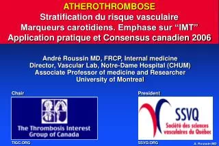
ATHEROTHROMBOSE Stratification du risque vasculaire Marqueurs carotidiens. Emphase sur “IMT” Application pratique et Consensus canadien 2006. André Roussin MD, FRCP, Internal medicine Director, Vascular Lab, Notre-Dame Hospital (CHUM) Associate Professor of medicine and Researcher

E N D
ATHEROTHROMBOSEStratification du risque vasculaireMarqueurs carotidiens. Emphase sur “IMT”Application pratique et Consensus canadien 2006 André Roussin MD, FRCP, Internal medicine Director, Vascular Lab, Notre-Dame Hospital (CHUM) Associate Professor of medicine and Researcher University of Montreal Chair President TIGC.ORG SSVQ.ORG
André Roussin MDDisclosures AstraZeneca Bristol-Myers Squibb Boeringher-Ingelheim GlaxoSmithKline Leo Pharma Merck Frosst Pfizer Roche Diagnostics Schering Canada sanofi aventis I have been on advisory boards or received honorarium as consultant or speaker or received research funds from the following companies:
HUMAN ATHEROGENESISFrom yellow streak to plaque and thrombosis 1 2 3 4 5 6 7 Libby P. Circulation. 2001;104:365-372
Inflammation markers Koenig W, Khuseyinova N. ATVB 2007; 27: 15-26
ASO and Drug Interventions Napoli C et al. Circulation 2006; 114: 2517-27
Cardiovascular disease worldwide • CVD (CAD, Stroke and PAD) is the leading cause of death worldwide1 • CVD contributed in 2001 nearly one third of all global deaths1-2 • 3 Risk factors are responsible for > 75% of all CVD worldwide1 • Elevated cholesterol • Smoking • High blood pressure • Of the three, elevated cholesterol carries the greatest attributable risk for CAD3 WHO. World Health report 2002 American Heart Association: statistical fact sheet 2003 Wilson P et al. Circ 1998; 97:1837-1847
Risque de développer MCAS pendant la vie 0.5 Femme 0.5 Homme 0.2 0.2 1/10 1/10 65 55 40 50 60 70 80 90 40 50 60 70 Age (années) Age (années) Lloyd-Jones, Lancet 1999; 353: 89-92
Notion « traditionnelle » de risque vasculaireConsensus Canadien sur les DyslipidémiesCalcul du risque de coronaropathie à 10 ans • ASO présente • Coronaropathie (MCAS) • Maladie artérielle périphérique • ASO carotidienne (ICT, AVC isch. , plaque) • Patients > 30 ans avec Diabète sucré • Dyslipidémie sévère • Hypercholestérolémie familiale (LDL) • Hypoalphalipoprotéinémie familiale (HDL) • Tous les autres • Préciser le risque avec les tables de Framingham du NCEP III Risque Élevé
Risque cardiovasculaire Framingham modifié NCEP IIIPour calculer le risque d’IM et demortalité CVPoints pour un homme 1. Age 2. Total Cholesterol (mmol/L) according to age
Risque cardiovasculaire Framingham modifié NCEP IIIPour calculer le risque d’IM et demortalité CVPoints pour un homme 3. Smoking according to age 5. Blood Pressure according to treatment 4. HDL-C
Pour calculer le risque d’IM et de mortalité CV Low Risk: < 10% Pour un homme Medium Risk: 10-20% High Risk: > 20%
INTERHEARTRisk of AMI associated with Risk Factors in the Overall PopulationODDS RATIO Yusuf S et al. Lancet 2004; 364: 937-52
INTERHEARTRisk of AMI associated with Risk Factors in the Overall PopulationPOPULATION ATTRIBUTABLE RISK Yusuf S et al. Lancet 2004; 364: 937-52
INTERHEARTRisk of AMI with Multiple Risk Factors 2.9 2.4 1.9 3.3 13.0 42.3 68.5 182.9 333.7 512 256 128 64 OR (99% CI) 32 16 8 4 2 1 Smk DM HTN ApoB/A 1+2+3 All 4 +Ob +PS All RFs Yusuf S et al. Lancet 2004; 364: 937-52
Notion « élargie » risque vasculaireIncluant le Consensus Canadien sur les DyslipidémiesAjoutant les facteurs de risque « émergents » • MCAS familiale précoce: RR = 1.7 à 2 • ApoB, Lp(a), LDL dense, ApoA1 • Syndrome métabolique • Marqueurs sub-cliniques d'ASO: • ITH, ECG effort, Plaques et Intima-media • Facteurs de risque émergents • hsCRP, homocystéine
Risk factors: markers and / or activators Atherosclerosis Atherothrombosis Stroke - MI - Death IM Plaque Stenosis Thrombosis Triggering Factors Smoking, Diabetes, LDL/oxLDL, HBP, AgII/AT1, Shear stress Endothelial Factors Inflammation Factors Cells, Intercellular + intracellular signaling, proteins-enz. actions Procoagulant Factors TF, PAI-1 / tPA and TxA2 / Prostacycline imbalances
New insights: What has been added Sub-clinical markers Ankle-Brachial Index Micro-albuminuria Carotid intima-media thick. Coronary calcification Serological markers hs-CRP Lipoprotein(a) Homocysteine Insulinemia sLp-PLA2 ++ +++ -/+ ++
CCS position statement 2006Treatment of dyslipidemia and prevention of CVD Adapté de: Can J Cardiol 2006; 22 (11): 913-927
Ultrasonographie carotidienneÉvaluation de l’ASO et stratification de risque CV Faible coût Accessible Non-invasive Imagerie excellente Quantitative Reproductible Mesure l’ASO intimale avant la sténose angiographique Épaisseur Intima-Media Intima-media thickness “IMT” Épaisseur de plaque Surface de plaque Volume de plaque Sténose Type de plaque: Échogénicité Homogénéité
Ultrasound Examination of the Carotid Artery External carotid Internal carotid Skin 1.0 cm Bifurcation 0.5-1.0 cm Commoncarotid 1.0 cm Near Wall Far Wall B-modeultrasound Periadventitia-adventitia Adventitia-media Intima-lumen Adventitia-periadventitia Media-adventitia Lumen-intima Smilde TJ et al. Lancet 2001; 357: 577-581
Façons de déterminer la valeur d’un marqueur de risqueVasan R S. Circ 2006; 113: 2335-2362
Considérations avant l’adoption d’un marqueur de risque CVVasan R S. Circ 2006; 113: 2335-2362
Marqueurs structurels et fonctionnels de risque CV Vasan R S. Circ 2006; 113: 2335-2362
Reproducibility of non-invasive ultrasonic measurement of carotid atherosclerosisThe Asymptomatic Carotid Artery Plaque Study (ACAPS) • 858 patients • 12 measurements in each patient • Repeated at 1 month • Within and between sonographer variation • Mean IMT difference (exam 2-exam 1) 0.13 mm • 90% of patients – mean difference < 0.2 mm Result • Highly reproducible measurement • B-mode ultrasound can monitor small rates of lesion progression Stroke 1992, Aug 23 (8), 1062-8
Protocoles pour Épaisseur Intima-Media (IMT) • 12 point manual measurement • Near and far wall of CCA, ICA, Bulb • Near and far wall of CCA, ICA • Far wall of CCA • Mean of maximal IMT measurement • Mean of mean IMT measurement • Manual VS automated edge detection • Plaque thickness summed • Plaque area summed • Plaque volume summed Adapted from Weingert M SSVQ 2006
IMTReproducibility of Measurement • Intra observer variability lower in studies limited to common carotid artery far wall (± 0.02 mm) VS multiple measurements at different carotid sites (± 0.06 mm) • Studies using automated computerized IMT measurement rather than manual cursor placement have best reproducibility. Adapted from Weingert M SSVQ 2006
IMT and ≥ 70% Coronary StenosisSensitivity vs Specificity IMT ofSensitivitySpecificity 0.6 mm 95% 20% 0.8 mm 55% 60% 1.0 mm 20% 90% Aminbaklish A. et al. Clin. Invest. Med 1999; 22:265-274
Evaluating Atherosclerosis by IMT measurementAnatomy 0.02 mm 0.80 mm Courtesy E. Braunwald Buithieu J /
Evaluating Atherosclerosis by IMT measurementMethodology • 12 point manual measurement • Far wall of Common Carotid Artery • Near and far wall of CCA, ICA • Near and far wall of CCA, ICA, Bulb • Mean of maximal IMT measurement • Mean of mean IMT measurement • Manual / automated edge detection • Summation of plaque thickness • Summation of plaque area • Summation of plaque volume ECA ICA ICA 10 mm Bulb 10 mm CCA 10 mm CCA Buithieu J /
Evaluating Atherosclerosis by computerized IMT measurement • ECG gating • Diastole • distal CCA • Mean IMT over 100 pts along at least 1 cm • Avoids pulsatile deformation of wall thickness • Observer independent • Better precision/reproducibility : Intermeasurement Δ = 3 % Automated Computerized method Buithieu J /
The Atherosclerosis Risk in Communities (ARIC) StudyPredictive Value of CIMT: Methodology • Prospective, multicenter study • N = 12841 aged 45 - 64 y (72.5 ± 5.5) • 7289 women, 5552 men • No evidence of CV disease at enrollment • Median follow-up 5.2 years • Mean CIMT over 1 cm - far walls of Right & Left CCA-Bulb-ICA ECA ICA 10 mm Bulb 10 mm CCA 10 mm Chambless LE & al. Am J Epidemiol 1997. 146:483-494 Buithieu J /
The Atherosclerosis Risk in Communities (ARIC) StudyPredictive Value of CIMT for Myocardial Infarct / Death Mean F-up 5.2 y Age and Gender adjusted CHD incidence/1000 patient-year CIMT (mm) Chambless LE & al. Am J Epidemiol 1997. 146:483-494 Buithieu J /
The Atherosclerosis Risk in Communities (ARIC) StudyPredictive Value of CIMT for Stroke Mean F-up 7.2 y Age and Gender adjusted Stroke incidence/1000 patient-year CIMT (mm) Chambless LE & al. Am J Epidemiol 2000. 151:478-487 Buithieu J /
The Atherosclerosis Risk in Communities (ARIC) StudyPredictive Value of CIMT by incremental value • CIMT (mean of CCA-Bulb-ICA) increment is associated with increased hazard rate ratio (HRR) Chambless LE & al. Am J Epidemiol 1997. 146:483-494 Chambless LE & al. Am J Epidemiol 2000. 151:478-487 Buithieu J /
The Atherosclerosis Risk in Communities (ARIC) StudyPredictive Value of CIMT by strata Hypertension 2.1 Diabetes 2.5 Current smoking 1.3 CIMT (mean of CCA-Bulb-ICA) increased hazard rate ratio (HRR) vs CIMT < 0.6 mm Chambless LE & al. Am J Epidemiol 1997. 146:483-494 Chambless LE & al. Am J Epidemiol 2000. 151:478-487 Buithieu J /
The Atherosclerosis Risk in Communities (ARIC) StudyPredictive Value of CIMT: Conclusions • N = 15 792 patients • CIMT measurements • Reproducible • Independent predictor of adverse cardiovascular eventsafter adjustment for: • Age, sex, race, center, BMI, waist-hip ratio, sporting activity • Diabetes, LDL, HDL, hypertension, smoking • Fibrinogen, WBC, LVH Chambless LE & al. Am J Epidemiol 1997. 146:483-494 Chambless LE & al. Am J Epidemiol 2000. 151:478-487
Predicting clinical coronary events: role of Carotid IMTCLAS Sub-Study • 133 patients: 8.8 year follow-up • Close correlation between far wall CCA-IMT and changes in catheterization • Progression of IMT correlated with: • Progression of CAD • Increased coronary events • Absolute IMT thickness and progression of IMT more strongly correlated with coronary events than • Changes in lipid levels • Lesion changes on coronary catheterization • Result: every 0.03 mm increase in IMT increases risk of coronary event 3.1 % Hodis H.N. et al Ann Int Med 1998; 128:262-269
Predicting clinical coronary events: role of Carotid IMTCLAS Sub-Study • CIMT directly associated withhigher risk for future MI and CHD death N = 146 CABG p < 0.001 CHD Risk Non fatal MI, Coronary Death, Revascularization Carotid Intima-Media Thickness (mm) Hodis HN & al. Ann Intern Med 1998. 128:262-269 Buithieu J /
Predicting clinical coronary events: role of Carotid IMT progressionCLAS Sub-Study • CIMT progression directly associated withhigher risk for future MI and CHD death N = 146 CABG p < 0.001 CHD Risk Non fatal MI, Coronary Death, Revascularization CIMT progression (mm/y) Hodis HN & al. Ann Intern Med 1998. 128:262-269 Buithieu J /
Cardiovascular Health Study (NHLBI)Predictive Value of CIMT: methodology • Prospective, multicenter study • N = 4476 aged > 65 y (72.5 ± 5.5) • Male 38.8 %, Caucasian 84.8 % • No evidence of CV disease at enrollment • Median follow-up 6.2 years • Maximal CIMT mean of near & far walls of R + L CCA • Maximal CIMT mean of near & far walls of R + L ICA O’Leary D & al N Eng J Med 1999;.340: 14-22 Buithieu J /
Cardiovascular Health Study (NHLBI)Predictive Value of CIMT for Myocardial Infarction & Stroke 100 95 1st Quintile 2nd Quintile 90 3rd Quintile 85 4th Quintile Cumulative Event-free Rate (%) 80 75 5th Quintile 0 1 6 7 0 2 3 4 5 Years 5 % 25 % O’Leary D & al N Eng J Med 1999;.340: 14-22 Buithieu J /
Cardiovascular Health Study (NHLBI)Predictive Value of CIMT for Myocardial Infarction & Stroke Myocardial Infarction or Stroke (Rate per 1000 Person-Years) Quintiles O’Leary D & al N Eng J Med 1999;.340: 14-22 Buithieu J /
Cardiovascular Health Study (NHLBI)Predictive Value of CIMT for Myocardial Infarction & Stroke * Relative Risk adjusted for age, sex, sBP, HTN, Atrial fibrillation, Diabetes O’Leary D & al N Eng J Med 1999;.340: 14-22 Buithieu J /
The Rotterdam StudyComparative Predictive Value for Incident Myocardial Infarction • Population-based cohort • N = 6389 aged > 55 (69.3 ± 9.2) • Male 38.1 %, Caucasian 100 % • No prior MI or revascularization • Mean Follow-up 4.2 years van der Meer IM & al. Circ 2004. 109:1089-1094
The Rotterdam StudyComparative Predictive Value for Incident Myocardial Infarction Composite atherosclerosis score • Carotid - Ultrasonography • Maximal CIMT mean of near and far wall of left & right CCA • Carotid plaque - weighted score • Aorta - Lateral abdominal X-ray • Calcifications - length of affected area • 0cm, <1.0, 1.0-2.5, 2.5-4.9, 5.0-9.9, ≥10.0cm • Lower extremities - Ankle-Brachial Index (ABI) • 1.50-1.21, 1.21-1.10, 1.10-0.97, 0.97-0.00 ? van der Meer IM & al. Circ 2004. 109:1089-1094
The Rotterdam StudyComparative Predictive Value for Incident Myocardial Infarction Adjusted HR Severity of Atherosclerosis None Mild Moderate Severe Carotid plaques 1.00 1.19 1.28 1.83 CIMT 1.00 1.56 1.63 1.95 Aortic Calcification 1.00 1.06 1.81 1.94 ABI 1.00 1.12 1.55 1.59 Composite Score 1.00 1.52 2.28 4.35 Incident MI : 258 / 6389 = 4.0 % van der Meer IM & al. Circ 2004. 109:1089-1094
Carotid PlaquePredictive value • 76 asymptomatic patients • Aged 35-65 • TC > 6.5 • Stress test, cath, carotid ultrasound ≥ 1 Plaque: 64% • 57% had critical CAD • Positive predictive value for coronary atherosclerosis: 76% No Plaque • Women: none had CAD • Men: - with positive stress test – 21% significant CAD Giral P. et al. Am J Card 1999; 84: 14-17
PLAQUE AREACAD rather than Stroke prediction Spence JD & al. Stroke 2002. 33(12):2910-2922 Buithieu J /