Vascular Surgery Back to Basics
Vascular Surgery Back to Basics. Dr. Tim Brandys Vascular and Endovascular Surgeon The Ottawa Hospital. OUTLINE. Acute limb ischemia Claudication Critical limb ischemia Aortic Aneurysm Aortic dissection Varicose veins Chronic venous insuffciency Superficial thrombophlebitis.

Vascular Surgery Back to Basics
E N D
Presentation Transcript
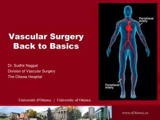
Vascular SurgeryBack to Basics Dr. Tim Brandys Vascular and Endovascular Surgeon The Ottawa Hospital
OUTLINE • Acute limb ischemia • Claudication • Critical limb ischemia • Aortic Aneurysm • Aortic dissection • Varicose veins • Chronic venous insuffciency • Superficial thrombophlebitis Acute Limb Ischemia
Mrs. Witelegg • ID: • 75 yo lady who lives by herself in an apartment. She is active, walks her dog ~2 kms daily without any difficulty. She takes pride in the fact that she has not needed to see a doctor in the last 10 years. • PMHx/PMSx: • remote TAH-BSO • social smoker quit in the 1960’s • no h/o DM, CAD, HTN, dyslipidemia, stroke, CRF Acute Limb Ischemia
Mrs. Witelegg • HPI: • While watching TV, she had sudden onset of numbness in her right leg. Her leg felt “like it went dead”, and she couldn’t ambulate. After a few minutes she experienced constant, severe pain starting in the toes, eventually involving the entire right leg. She called her neighbor and then brought her in to the Civic emergency department. Acute Limb Ischemia
Mrs. Witelegg • What is Acute limb Ischemia? • An abrupt cessation of arterial blood flow to an extremity resulting in hypoperfusion of tissue, threatening limb viability Acute Limb Ischemia
Mrs. Witelegg • O/E • She is in distress from pain in R leg • BP:140/90 mmHg, HR:150 bpm • pulse: irregularly irregular • Normal heart sounds, good a/e bilat • No pulsatile masses in her abdomen • No carotid, abdominal or femoral bruits • Pulses: • L: + femoral, + politeal, + DP, + PT • R: - femoral, - popliteal, - DP, - PT • R foot is colder and paler than L • Decreased sensation in R foot • Able to move toes but difficulty with plantar and dorsi flexion • Absence of trophic changes in her lower extremities (no hair loss, thickened nails, or thin, flaky or shiny skin) Acute Limb Ischemia
Mrs. Witelegg • What are the 6 P’s of Acute Limb Ischemia • Pain • Palor • Polar/poikilothermia • Paraesthesia • Paralysis • Pulselessness Acute Limb Ischemia
Mrs. Witelegg • Classify Acute Limb Ischemia. In which category is Mrs. Witelegg? Acute Limb Ischemia
Mrs. Witelegg • Your working diagnosis is acute limb ischemia. • You order CBC, electrolytes, BUN, Cr, PTT/INR (all of which comes back normal), type and cross-match blood, and a saline infusion is started. • CXR is unremarkable • ECG is as follows: Acute Limb Ischemia
Mrs. Witelegg • Before you call the vascular surgeon on-call, what test can you do at the bed side that can objectively assess acute limb ischemia? • Ankle Brachial index • Measure brachial pressure (example 160 mmHg) • Measure ankle pressure (example: 80 mmHg) • Divide ankle by brachial pressure (exmple 80/160 = 0.5; anything <0.9 is abnormal) Acute Limb Ischemia
Mrs. Witelegg • What is the most likely etiology of ALI in Mrs. Witelegg? • Cardiogenic embolism • What in her history and physical supports this diagnosis? • Lack of atherosclerotic risk factors • no previous claudication (she walked her dog 2 km/day) • Irregularly irregular pulse • Completely normal left extremity pulses • Based on her physical examination, what is the highest point of obstruction of arterial flow? • R ileo-femoral region • How long can a limb be without blood flow before irreversible tissue damage ensues? • 4-6 hrs Acute Limb Ischemia
Mrs. Witelegg • What is the surgical management of this condition? • R femoral embolectomy • Can we proceed to the OR without any imaging studies? If not what studies can be perfomed? • Because of the classic history and physical findings, and because of the presence of class 2b ischemia, immediate surgery is indicated without delay for imaging. • Angiography can be performed in certain conditions of ALI • when the suspected etiology is arterial thrombosis (i.e. in preparation for bypass surgery) • when the patient has class 1 or 2a ischemia Acute Limb Ischemia
Mrs. Witelegg • What medical therpay is available for ALI and when is it indicated? • Lytic therapy (i.e. with t-PA) is used to dissolve the clot. It is a good option in the setting of acute arterial or graft thrombosis. It is not indicated in the setting of trauma or when the patient can not wait more than 24-48 hrs, as the therapy requires that period of time for clot dissolution. ( i.e. class 1 or early 2a ischemia) • IV Heparin will not dissolve the clot but will prevent further propagation, and is only indicated if there is a delay to surgery
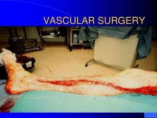
Mrs. Witelegg • The patient is booked for emergency embolectomy • Under local anaesthesia, a small incision is made over the R groin. The femoral artery is exposed and controlled with vessel loops. A small arteriotomy is made and the clot is removed proximally and distally using a fogarty balloon embolectomy catheter. • The arteriotomy is repaired and the foots “pinks up” after blood flow is returned. There is a palpable DP and PT pulse. • The patient is returned to the recovery room. Acute Limb Ischemia
Mrs. Witelegg • At 3 am you get paged by the recovery room nurse. Mrs. W is complaining of significant pain in her leg, it is more swollen and the DP and PT are no longer palpable. • In addition, her urine output has diminished and she is peeing out dark urine which tested positive for “blood” on the urine dipstick. Acute Limb Ischemia
Mrs. Witelegg • What is happening to Mrs. W? • Reperfusion syndrome: occurs as a result of blood flow going back into previously damaged tissue, causing rhabdomyolysis and compartment syndrome.. • Rhabdomyolysis: Liberated myoglobin from dead muscle cells enters the blood stream resulting in renal tubular obstruction and direct nephrotoxicity causing renal failure. Myoglobinuria is a false positive on the urine dipstick test for blood. • Compartment syndrome: Free oxygen radicals are created with reperfusion. These result in increased tissue edema, with in the limited facial compartments of the lower leg, this further decreases capillary blood flow and worsens the ischemia and tissue damage, causing further edema. Pain out of proportion, pain on passive stretch and high pressures in the compartments suggests compartment syndrome. Acute Limb Ischemia
Mrs. Witelegg • How should reperfusion syndrome be managed? • Compartment syndrome is a surgical emergency and is managed by 4-compartment fasciotomies. • Rhabdomyolysis should be managed with aggressive IV fluids, diuresis and alkalinization of urine. Acute Limb Ischemia
AORTIC DISSECTION Vascular Surgery – Back to Basics
Definition • spontaneous tear in aortic intima allowing blood to be driven between the aortic intima and media • acute < 2 weeks • chronic > 2 weeks
Classification • DeBakey • Type I - involves ascending and descending aorta • Type II - ascending aorta only • Type IIIA - descending thoracic aorta • Type IIIB - Type IIIA plus abdominal aorta • Standford • Type A - ascending aorta and aortic arch; emergency • Type B - aorta distal to subclavian artery; emergency surgery if complications of dissection
Etiology • HYPERTENSION, usually uncontrolled • TRAUMA, usually deceleration injury (falls, MVAs) • other: cystic medial necrosis, atherosclerosis, connective tissue disease (Marfan’s syndrome, Ehlers-Danlos syndromes), congenital conditions (coarctation of aorta, bicuspid aortic valves, PDA), infection, arteritis (Takayasu’s)
Epidemiology • incidence 5.2 in 1,000,000 • male:female = 3:1 • small increased incidence in African-Canadians (related to higher incidence of hypertension) • lowest incidence in Asians
Clinical Features • SUDDEN ONSET SEVERE CHEST PAIN RADIATION TO THE BACK (INTERSCAPULAR) +/-.... • hypertension • asymmetric BP’s and pulses between arms • ischemic syndromes due to occlusion of aortic branches: coronary (MI), carotid (stroke, Horner’s syndrome), splanchnic (ischemic gut), renal (kidney failure) • “unseating” of aortic valve cusps (new diastolic murmur) • rupture into pleura (dyspnea, hemoptysis) or peritoneum (hypotension, shock) or pericardium (tamponade) • lower limb ischemia (cold legs)
Investigations • CT scan is gold standard • CXR • pleural cap • widened mediastinum • left pleural effusion with extravasation of blood • TEE • ECG: LVH (90%), +/- MI, pericarditis, heart block • aortography, MRI
Treatment • Type A • EMERGENCY CARDIAC SURGERY • may require putting patient on pump, hypothermic circulatory arrest, valve replacement, coronary re-implantation of aortic root • resection of intimal tear, reconstitution of flow through true lumen, replacement of the affected aorta with graft • Type B • MEDICAL MANAGEMENT • very rarely urgent operation for complications (expansion, rupture, gut/leg/renal ischemia, ongoing pain
Definition • localized dilation of an artery that is 2 x its normal diameter • true aneurysm: involving all vessel wall layers • false aneurysm: disruption of aortic wall with containment of blood by some layers of the aorta or a fibrous capsule made of surrounding tissue
Etiology • DEGENERATIVE (matrix metalloproteinases) • atherosclerosis association • infection • cystic medial necrosis • trauma • vascultitis • connective tissue disease (Marfan syndrome, Ehlers-Danlos)
Epidemiology • incidence 5 to 32 per 100,000 for AAA • high risk groups: • 65 years and older • male:female = 4:1 • smokers • peripheral vascular disease, CAD, CVD • family history of AAA
Clinical Features • Vast majority ASYMPTOMATIC • RUPTURE • back pain • hypotension/syncope • pulsatile abdominal mass • ~100% mortality if untreated
Investigations • abdominal US (100% sensitive) • CT • Aortogram (false negative normal lumen size due to thrombus formation)
Treatment • Risk of rupture depends on size • <5 cm <5% / yr • 5-6 cm 10% / yr • 6-7 cm 15-20% / yr • >7 cm >20% / yr • Risk of dying from aneurysm surgery = ~5%
Treatment • Operate when • AAA reaches 5.5 cm in an otherwise healthy individual • >5 mm expansion in 6 months • symptomatic AAA • Rupture • contraindications: life expectancy < 1 year, terminal disease (cancer), significant co-morbidities (recent MI, unstable angina), severe dementia, advanced age
Treatment: Surgical • Surgical options: • open surgery with graft replacement • Endovascular aneurysm repair
Ruptured Aortic Aneurysm • EMERGENCY • clinical diagnosis class diagnostic triad (50% cases) • sudden onset back pain • shock (syncope/hypotension) • pulsatile mass • U/S in emerg or CT if stable • IV access, start fluid resuscitation, cross and match • EMERGENCY LAPAROTOMY and CLAMP AORTA • Prognosis • 100% mortality untreated, OR mortality rate 50%; 90%total mortality
Clinical Features - Claudication • Pain with exertion (usually calves) • relieved by short rest - two to five minutes • reproducible • P/E • hair loss, hypertrophic nails, atrophic muscle • pulses may be absent at some locations
Etiology • blockages in arteries to lower extremities due to atherosclerosis • Risk factors • smoking • DM • HTN • hyperlipidemia • family history • obesity • sedentary • male gender
Investigations • Ankle Brachial Index • Angiogram
Treatment • CONSERVATIVE • risk factor modification • exercise program • cilostazol • anti platelet (ECASA, clopidrogel) for MI / stroke risk • surgical • indications: claudication interfering with lifestyle • options: endovascular, PTA, arterial bypass grafts
Clinical Features - Critical limb ischemia • Pain at rest in foot, worse at night • relieved by short rest - two to five minutes • reproducible • pulses may be absent at some locations • P/E • hair loss, hypertrophic nails, atrophic muscle • ruborous foot • ulcers • gangrene
Etiology • blockages in arteries to lower extremities due to atherosclerosis • Risk factors • smoking • DM • HTN • hyperlipidemia • family history • obesity • sedentary • male gender
Investigations • Ankle Brachial Index • Duplex ultrasound • Angiogram
Treatment • Surgical • bypass • gortex vs vein • Endovascular balloon angioplasty • limited durability • less morbid