VASCULAR SURGERY
VASCULAR SURGERY. Cerebrovascular Disease. CAROTID:. Presentation : Asymptomatic : Bruit (only 20% hemodynamically significant lesion) Screening prior to other surgery. Presentation:. Symptomatic: TIA, Stroke Amaurosis fugax ipsilateral to carotid lesion

VASCULAR SURGERY
E N D
Presentation Transcript
CAROTID: Presentation: • Asymptomatic: • Bruit (only 20% hemodynamically significant lesion) • Screening prior to other surgery
Presentation: • Symptomatic: • TIA, Stroke • Amaurosis fugax ipsilateral to carotid lesion • Contralateral motor or sensory deficit • Facial droop • Dyshasia or aphasia
Investigations • Duplex Scan • CT scan - confirm or r/o infarct • CT/Angio – Confirm U/S plan OR • MRA – Similar to CT
Management • Asymptomatic - Risk factor reduction (asa,statin,ACE) • observation with regular duplex scans • Antiplatelet agent and surgery more controversial • ACAS 60% OR • Canada 80% male, under 75 yrs or operate
Symptomatic: Carotid Stenosis 70% TIA, Small completed stroke with minimal residual neurologic deficit, antiplatelet agent +carotid endarterectomy
Arterial Aneurysms • Definition:1.5-2x diameter adjacent normal artery. • Ex. Aorta 3 cm • True: All layers of arterial wall dilated • False: Aneurysm usually consists of hematoma +/- adventitia
Distribution of Aneurysms • Aorta: 90-95% are infrarenal • Peripheral: Popliteal most common, 2nd femoral • Visceral: uncommon, splenic (most common)
Aortic Aneurysms • Risk factors: male, age 60 yrs, smoking, COPD, FHx +ve, CAD, PVD, peripheral aneurysms. • Natural Hx: AAA 5 cm grow 0.3-0.5 cm/year • Rupture rate: • 5 cm - 1.5% over 5 yrs • 5.5-5.9 cm - 25% over 1-5 yrs • 6 cm - 35% over 1-5 yrs • > 7 cm - > 75% over 1-5 yrs
AAA Presentation • Asymptomatic: incidental finding on Px or Radiologic Test • Symptomatic: ABD/BACK pain (leak or rapid expanding) • Rupture: 35% initial presentation, Triad ABD/BACK pain, Hypotension, Pulsatile Mass.
AAA Detection • Physical Exam - not sensitive • U/S ABD - highly sensitive and specific • CT / MRI - sensitive, specific, but expensive • Angio - not reliable
AAA Management Indications for Surgery: • risk of rupture > surgical risk • size 5 cm FEMALE • > 5.5 cm MALE • symptomatic • ruptured • rapid expansion Observation with U/S q6 months if asymptomatic and < 5 cm.
Acute Limb Ischemia • Sudden onset of sxs/signs • Severity presentation depends on adequacy of collateral circulation • 5 or 6 P’s: pain, pallor, pulselessness, paralysis, paresthesia, +/- poikilothermia
CAUSES • Embolus • Thrombosis • Trauma
Embolus • Clot displaced from site of origin to occlude a distant artery • Most common site to lodge bifurcation common femoral artery • 90% come from the heart (atrial fibrillation, recent M.I.)
Thrombosis • Clot forms in situ in a previously diseased vessel or bypass graft • Predisposing factors: Dehydration, CHF or Hypercoagulable state
Embolus Dramatic presentation (sudden onset) Opposite leg normal pulses Source for embolus: A.fib, recent M.I. Thrombosis Bland (well dev. collaterals Opposite leg abn. Pulses Hx of chronic PVD, ex. claudication Acute Arterial Occlusion Presentation
Investigations • Angiogram/CTA • Gold Std • Embolus (not always needed prior to OR but shows abrupt cut off of circulation, reverse meniscus sign, no collaterals. • Thrombosis - always needed, shows tapering cut off, lots of collaterals
Treatment Embolus: • Anticoagulate with Heparin • Medical resuscitation • Surgical embolectomy • Consider Fasciotomies • Post-op life long anticoagulation Heparin Coumadin
Treatment • Thrombosis • angiogram always • thrombolysis +/- later surgical intervention • Endovascular (angioplasty/stent) • surgical bypass • post-op antiplatelet agents
Compartment Syndrome • Especially after reperfusion of the leg • pressure within fascial compartments >30mmHg. • Symptoms/signs: Pain out of proportion, pain on passive flexion/extension, absent pulses is a very late sign • Treat: fasciotomies
Presentation (symptoms): • Claudication = Reproducible pain in the lower extremities on ambulation • Rest pain = Pain at rest in forefoot, toes. Constant pain, worse at nite
Presentation (signs): • Claudicant +/- pulse deficits • Rest pain - pulse deficits, atrophic skin, hair loss on toes • Tissue loss - ulcers (painful), gangrene
Presentation (signs): • Ankle brachial index: • Normal 1 • Claudication 0.5 - 0.8 • Rest pain < 0.5 • Tissue loss < 0.3 • ABI not always reliable in diabetic patient • Doppler signal present does not always ensure adequate circulation
Leriche Syndrome: • Absent femoral pulses • Impotence • Buttock Claudication
Investigations: • Hx, Px, ABI • Blood Flow Lab - Duplex scan, exercise testing, segmental pressure studies • Angiogram/CTA - Indicated prior to intervention or diagnostic dilemma
Conservative Management • Modify risk factors - smoking cessation, hyperlipidemia, diabetes • Walking exercise program (Develops collateral circulation) • MEDS: • All should be on antiplatelet: ECASA, Clopidogrel, Ticlopidine, etc • Statin • Consider Pentoxifylline
INTERVENTION • Indications • disabling claudication • Critical ischemia:rest pain, tissue loss • Angioplasty + Stenting (best results for proximal lesions ex: iliac lesion) • Bypass • Amputation
Aortic dissection • Definition: Intimal tear leading to creation of a false passage way of blood within the wall of the aorta. Results in both a true and false lumen of the vessel
AORTIC DISSECTON • Most common catastrophic event of the aorta • Consequences include: • weakening of aortic wall and possible rupture • interruption of blood supply to branches of the aorta involved, resulting in end organ or limb ischemia
Presentation • Classically older patient with hx HTN and sudden onset “tearing” retrosternal chest + back pain • On examination: HTN, pulse deficits are possible, murmur of aortic regurgitation
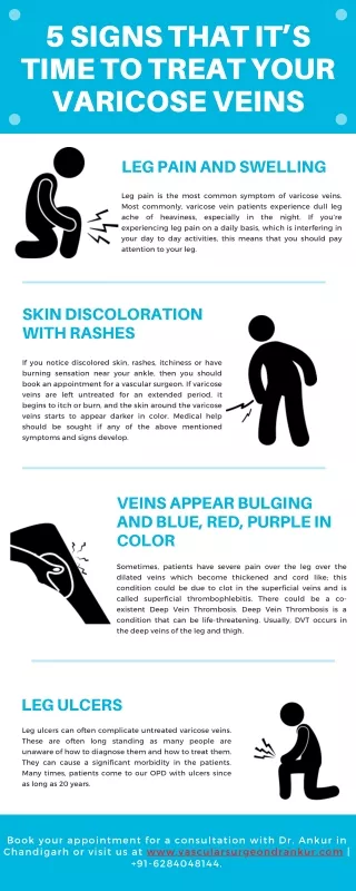
Varicose Veins • Dilated saccular or cylindrical superficial veins • Different appearances/severities • Telangiectasia (spider cluster extending out from feeder vessel). • Stem veins (saphenous) • Reticular veins (tributaries).
Classification • Primary - Superficial venous system only • Secondary - Deep system and or perforators are also abnormal usually as result of DVT, Pregnancy, Trauma
Predisposing Factors • Family history, female, 50 yrs or older, multiparity, standing occupation, obesity, BCP, DVT
Pathophysiology Primary Varicose Veins • Controversial: valvular incompetence, wall weakness, A-V fistula
Presentation (symptoms) • Cosmetic appearance • Pain, leg fatigue, burning, itching • Swelling • Symptoms made worse by prolonged standing, relieved with elevation