PERIPHERAL VASCULAR SURGERY
PERIPHERAL VASCULAR SURGERY. Summary. Anatomy & Physiology Pathology Diagnostic Exams Preparation Prep/Positioning Basic Supplies, Equipment, Instrumentation Peripheral Vascular Procedures: Vascular access Carotid endarterectomy Bypass procedures. Terminology .


PERIPHERAL VASCULAR SURGERY
E N D
Presentation Transcript
Summary • Anatomy & Physiology • Pathology • Diagnostic Exams • Preparation Prep/Positioning • Basic Supplies, Equipment, Instrumentation • Peripheral Vascular Procedures: • Vascular access • Carotid endarterectomy • Bypass procedures
Terminology • Arrhythmia-irregular heart rhythm • Arteriosclerosis-hardening of the arteries (part of aging process) • Atherosclerosis-build-up of plaque • Autogenous/autologous-originates in the body • Bifurcation-fork/point of branching • Cannula-tube/sheath allowing passage of fluids • Cardiopulmonary-r/t heart and lungs • Claudication-cramping, aching, stiffness caused by exercise relieved by rest (1° sx. PVD) • Cyanosis-blue discoloration of an extremity or the skin caused by lack of oxygenation (Hgb) • Embolus-matter traveling through a vessel • Extracorporeal-outside the body • Fibrillation-rapid, ineffectual contractions of the heart • Defibrillation-to stop fibrillation by drugs or electrical means • Lumen-space within an artery, vein or tube
Terminology Continued • Occlusion-abnormal obstruction/closure of a vessel • Palliative-to relieve without curing • Plaque-patch of atheromatous matter (cholesterol, lipids, cellular debris) that forms in the inner lining of an artery (intimal lining) • PVC (premature ventricular contraction)-arrhythmia that precedes normal electrical impulse/may precede ventricular fibrillation • Septum-wall that separates two cavities • Stenosis-narrowing or constriction of a vessel • Thrombus-blood clot (thrombus) • TIA (transient ischemic attack)-temporary interference of brain oxygenation by the arteries Symptoms may last a few minutes to several hours • Vasoconstriction-narrowing of a vessel
The Peripheral Vascular System • A closed system of the body that carries blood from the left side of the heart that has been oxygenated in the lungs→ to the heart itself, all organs, and tissues of the body where the oxygen is utilized→ back to the right side of the heart where it will be sent back to the lungs for re-oxygenation to start the cycle over again
Peripheral Vascular System Composition • Two Types of VESSELS: • Arteries • Veins
VESSELS(Arteries) • Arterial blood is pumped from the heart to the rest of the body via vessels called arteries • Arterial blood is going away from the heart • Arteries are large vessels originating with the AORTA that come directly out of the heart • Arteries divide into smaller braches as they reach their destination in the body • Arteries→arterioles→capillaries
Capillaries • Microscopic level of: • oxygen & carbon dioxide exchange • nutrient exchange • waste exchange between blood and tissue fluid in areas called capillary beds
Venules • Capillaries join the smallest veins called venules which become larger in size to become veins which ultimately end at the superior vena cava and inferior vena cava in the right atria of the heart where unoxygenated blood is sent back to the lungs via the pulmonary artery for reoxygenation
VESSELS(Veins) • Veins take blood back to the heart for reoxygenation • Capillary bed→Venules→Veins→Vena Cava (Superior and Inferior)
Vessel Structure • 3 layers called tunics • Inner = tunica intima • Middle = tunica media • Outer = tunica adventitia
Differences in Vessel Structure(Arterial) • Tunica Intima • Inner tunic has an endothelium lining • Smooth layer that is in contact with blood to promote flow and prevent damage to the platelets
Differences in Vessel Structure(Arterial) • Tunica media • thickest layer • layer of smooth muscle can contract or dilate with autonomic nervous system impulses • contraction = vasoconstriction = ↑ BP • dilation = vasodilation = ↓ BP
Differences in Vessel Structure(Arterial) • Tunica Adventitia • Outer tunic • Consists of connective tissue that connects arteries to tissues that surround them • Contains vaso vasorum which are vessels that nourish the arterial wall
Differences in Vessel Structure(Veins) • Same three layers as arteries • Differences are in the thickness of each layer • Tunica adventitia is thickest layer • Tunica media has less smooth muscle tissue than arteries • Tunica intima is thinner than an artery and contains valves • Vein lumen is larger than an artery lumen
Blood Pressure • Force blood exerts on the inner walls of vessels as it passes through them • Veins: • Low pressure • Working against gravity • Movement by skeletal muscle contraction as blood moves up to the heart (Veins are surrounded by skeletal muscle) • Backflow prevented by valves in the veins
Blood Pressure • Arteries: • High pressure • Dependent On: • Volume • Ventricular contraction strength • Resistance • Viscosity (thickness) • Heart rate
Blood Pressure • Systole = contraction • Diastole = relaxation • Central Venous Pressure = venous blood pressure in the right atrium measured with a central venous catheter (normal is 3-8)
Blood Flow • Blood that travels undisturbed through the vessel is called laminar • Blood that is disturbed by an obstruction, stenosis, curve, or bifurcation is called turbulent • Turbulence can be auscultated by doppler and is called a bruit • Turbulence that can be felt or palpated is called a thrill
Arterial System • Ascending Aorta→coronaries • Aortic Arch: 3major branches • First branch= brachiocephalic (innominate) • Brachiocephalic bifurcates into right subclavian and right common carotid • Second branch=left common carotid • Third branch=left subclavian • Descending Aorta: • Above the diaphragm, aorta = thoracic aorta • Below the diaphragm aorta = abdominal aorta
Upper Extremities (arterial) • Right subclavian>right arm>axillary artery>brachial artery>bifurcates to form ulner and radial arteries>rejoin at palmer digital arteries • Left subclavian>left arm>axillary artery>brachial artery>bifurcates to form ulnar and radial arteries>rejoin at palmer digital arteries
Head (arterial) • Right common carotid and left common carotid > brain, head, and neck • Common carotids bifurcate to form internal and external carotid arteries • External carotids>neck and head • Internal carotids>join vertebral artery (off subclavian) to form basilar artery >form Circle of Willis in the brain
Abdominal Aorta • Supplies oxygenated blood to the abdominal wall and abdominal organs/viscera
Lower Extremities (arterial) • Aorta bifurcates to form right and left common iliac arteries • Common iliacs bifurcate to form internal and external iliacs • Internal iliacs supply pelvis and perineum • External iliacs become femoral arteries>popliteal>bifurcates to form anterior tibial and posterior tibial • Anterior tibial becomes dorsalis pedis>plantar arch arteries • Posterior tibial>peroneal artery>joins dorsalis pedis to form plantar arch arteries
Venous System • Internal jugular veins drain the brain, head, face, and neck> subclavian veins> this union is called the innominate or brachiocephalic vein • Leads to the Superior Vena Cava which empties into the right atrium • External jugulars drain parotid glands and the superficial face and scalp> subclavian veins>SVC • Vertebral veins drain neck and vertebrae>subclavian veins>SVC
Venous System Continued • Upper Extremities (superficially)are drained by the basilic and cephalic veins that empty into axillary vein>the subclavians>SVC • Upper Extremities (deep) are drained by the radial, ulnar, and brachial veins>axillary vein>subclavians>SVC
Venous System Continued • Lower Body drains via those veins into the Inferior Vena Cava which also empties into the right atrium • See Overhead
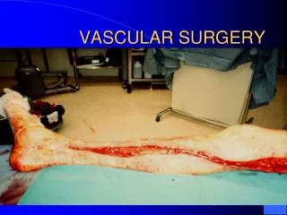
Arterial Insufficiency (2 types): 1.Acute Embolic or an unstable atherosclerotic plaque rupturing and creating a thrombosis or clot 80% in lower extremities Definition/Clarification: Embolus is a foreign substance or blood clot (liquid, solid, or gas) transported by the blood or lymphatic system ex. clot, air, fat, tumor parts Thrombosis is a blood clot that occludes a vessel If detached it becomes an embolus Emboli usually come from the heart during an MI or A-Fib, can come from other areas and attach itself (usually attaches at bifurcations or narrowing areas) Creates loss of circulation to areas below it S/SX:5 Ps (pulselessness, pallor, pain, parethesia, and paralysis) Arterial Disease
Acute Arterial Insufficiency Continued • Can patient tolerate arteriograms and anesthesia • Medical intervention is choice with unstable patient (thrombolytics) • Surgical intervention when stable=arterial embolectomy • Limb not salvageable=amputation
2. Chronic = Ischemia Results in inhibited or total blockage of flow 2 types: a. Arteriosclerosis Arteriosclerosis is part of the aging process creates hardening of the arteries= less elastic Atheroma=thickening of tunica intima seen with arteriosclerosis b. Atherosclerosis Atherosclerosis is this build-up of plaque Result of calcium or cholesterol deposits (plaque) inside the tunica intima Arterial Insufficiency
Atherosclerosis • Gradual process • Body develops collateral circulation as a compensatory mechanism • Causes speculated as intimal damage from smoking, hypertension, diabetes, etc. • Often referred to as atherosclerosis obliterens
Atherosclerosis • Generally is segmental in occurrence which allows for surgical intervention to correct it • If not corrected, can lead to gangrene or tissue death below the blockage in extremities • In the carotid arteries can lead to stroke • Surgical intervention involves bypass grafting (native vein or graft material) or endarterectomy (removal of plaque)
Aneurysms (peripheral) • Trueaneurysm=dilation of all layers of the arterial wall • May find atherosclerosis along with true aneurysm/is not the cause of • False Aneurysm (pseudoaneurysm)=not an aneurysm, but a tear that allows blood between the layers of the artery • Results from trauma, infection or post-arterial surgery where suture has been disrupted
Venous Insufficiency • Caused by deep venous thrombosis • Results from injury to the endothelium of the vein, stasis (immobility), coagulapathy problems, orthopedic trauma • Usually lower extremity clot • Urgent situation as clot can dislodge and move into the right atrium and make its way to the pulmonary artery resulting in death (PE=pulmonary embolus) • Medical treatment= anticoagulants • Can do a thrombectomy if isolated • Long term=vena cava filter
Diagnostic Exams • Angiography = Gold Standard for diagnosis with peripheral vascular disease • Ultrasound-detection by sound waves • Doppler-Measures blood flow • Computed Axial Tomography (CAT/CT Scan)-x-ray pictures in slices • Magnetic Resonance Imaging (MRI)-uses radio waves and a magnetic field to provide the 3-D views (can move in any direction unlike CT and is nonradioactive)
Anesthesia • Patient dependent: general, spinal, epidural, or local • All spinal/epidural patients get a foley catheter • CAE: will use an EEG to monitor brain activity and determine if a shunt is needed during the procedure. Can be done by CRNA or an EEG technician
Medications • Saline with antibiotic irrigant of surgeon choice or one patient is not allergic to • Heparin saline or lactated ringer’s irrigation for washing out inside artery to prevent clot during surgery (usually 250ml NS to 1,000units Heparin) • Papaverine antispasmodic/smooth muscle relaxant 120mg to 250ml NS (distention, prep, storage of vein grafts) • Topical Hemostatic Agents: Surgicel, Gelfoam with Thrombin, Avitene, other fibrins (floseal, tisseal) (Surgeon choice)
Positioning • Extreme Care Taken with Positioning due to limited Circulation of these Patients • Try to position while awake to get feedback from patient • Pay attention to anatomical alignment • Padding bony prominences • DO NOT lay heavy instruments on patient • Supine with arms tucked or on armboards • Pillow under knees • Pads under heels and arms • Pillow, headrest, or donut under head (avoid neck hyperextension) • Shoulder roll for neck extension needed for carotid endarterectomy
Prep (Considerations) • Doctor preference/Patient allergy: Hibiclens, Betadine • Non-open wounds an Ioban is preferred due to fact that are operating on vasculature which is a potential opening to septicemia • If scrubbing a carotid or aneurysm BE GENTLE! You could loosen plaque or rupture an already ready to rupture artery!
Preps • Extensive/Circumferential • Nipples to knees for AAA (flat) • Pubis to ankle or whole foot (lower extremity) • May be from the waist down if using vein graft from one leg to the other • CAE ear lobe of affected side to clavicle/maybe to nipple and well across the chest. Head should be turned to expose affected side and a shoulder roll may be needed to provide a smooth surface
Drapes IMPERVIOUS DRAPES • Extremity drapes • Universal drapes • Pediatric Laparotomy sheet • U-sheet
Basic Supplies, Equipment, Instrumentation • Drape Pack Clips • Minor or Major basin Rubber shods • Specialty Trays (CV or PV) Contrast • Vessel loops/umbilical tapes Kittner/peanut • Heparin needle or angiocath Tunneler • Silk ties or reels Introducer kit (prn) • Vessel suture: Prolene or Surgilene • Drain suture: nylon or Ethilon • Subcuticular suture: Vicryl or Dexon • Subcutaneous layer: staples, Ethilon, Monocryl, Vicryl, or Dexon