Download
1 / 11
110 likes | 241 Vues
Enhanced Wide Angle (EWA) Forward Light Scatter Improves Resolution of Red Blood Cell Microvesicles (RBCMV). E. Michael Meyer 1 , Chenell L. Donadee 2 , Mark T. Gladwin 2 , Albert D. Donnenberg 1,3

E N D
Enhanced Wide Angle (EWA) Forward Light Scatter Improves Resolution of Red Blood Cell Microvesicles (RBCMV) E. Michael Meyer1, Chenell L. Donadee2, Mark T. Gladwin2, Albert D. Donnenberg1,3 University of Pittsburgh Cancer Institue Cytometry Facility, University of Pittsburgh, Pittsburgh, PA1 Division of Pulmonary Allergy and Critical Care Medicine University of Pittsburgh Medical Center, Pittsburgh PA2 University of Pittsburgh School of Medicine, Department of Medicine, Pittsburgh PA3
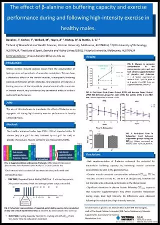
Abstract Detection of red blood cell (RBC) derived microvesicles is an area of intense interest because they hold promise as a biomarker of disease activity in sickle cell anemia, thalassaemia intermedia and cardiovascular disease. Their quantification in stored blood products may also serve as an objective quality indicator. Flow cytometry based assays have been devised to quantify RBCMV in blood on the basis of conventional forward light scatter (FSC) and annexin V binding. In such assays, RBCMV are detected as a discrete population of FSC low cells, the majority of which bind annexin V, indicating loss of phospholipid asymmetry. Intact RBC are largely annexin negative, but form one continuous population with transitional cells having slightly lower FSC and higher annexin binding. Presumably this transitional population is intermediate to the formation of RBCMV. The newly released Beckman-Coulter Gallios cytometer has the capability to detect FSC light in a mode that preferentially amplifies signals collected between 8-19º. This mode, termed Enhanced Wide Angle Forward Scatter, is particularly useful for resolving size difference among small particles (micron to submicron range). In this study, we demonstrate that EWA FSC is able to resolve RBCMV from both the transitional population and from intact RBC in blood bank whole blood products that had been aged 3 and 21 days. Intact RBC and fragments were detected as Glycophorin-A (Gly-A, CD235a) (PE-Cy5) positive events. Annexin V binding was measured on each Gly-A+ population. With EWA FSC, intact RBCs were detected as a homogenous scatter population with a mean half-peak CV of 10.8%. This population was easily distinguished from the lower scatter transitional population and from microvesicles themselves. In contrast, using conventional FSC, RBCs formed a diffuse population (mean half-peak CV = 17.7%) that was continuous with transitional vesicles. The ability to resolve RBC populations by EWA FSC enables further time course studies to determine whether the emergence of a transitional population predicts the subsequent degradation of RBC into RBCMV.
Beckman-Coulter Gallios: Forward Scatter Collection in Three Modes Select From: Low Angle Light Scatter Wide Angle Light Scatter Enhanced Wide Angle Light Scatter Enhanced Wide Angle Light Scatter increases sensitivity without increasing noise. The Gallios’ photo-diode has a sophisticated design that allows it to amplify wide angle light scatter independently of low angle light scatter. This option has allowed us to study small particles such as microvesicles resulting from red blood cell hemolysis. This feature could be beneficial to anybody in need of enhanced sensitivity when detecting small particles.
Microvesicles as a Function of Blood Age 16.00% 12.00% 8.00% Percent Microvesicle 4.00% 0.00% 0 10 20 30 40 Days in Storage Stored Blood: Microvesicles Increase versus Time To detect microvesicles in blood product, an assay has been developed that requires less than 1ml of sample. Detection of microvesicles could prove to be a good indicator of expired blood products. Increasing evidence has suggested that increased levels of microvesicles in blood products could have severe physiological consequences. Rubin, et.al Vox Sanguinis (2008) 95, 288-297
Microvesicles are Part of a Bigger Picture Rother. JAMA. 2005.
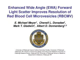
Hemolysis Effects NO Levels Blood NO in physiology, pathology, and therapeutics. Red blood cells (RBCs) are shown in a blood vessel where the endothelium containing nitric oxide synthase (NOS) is surrounded by smooth muscle cells that contain soluble guanylyl cyclase (sGC enzyme; not shown). The factors involved in reducing RBC scavenging of NO are the cell free zone, the unstirred layer, and the intrinsic membrane barrier. NO formation in the RBC is accomplished by both the RBC NO Synthase and deoxy hemoglobin mediated reduction of nitrite. The RBC in the middle illustrates the pathology associated with hemolytic conditions and microvesicle formation proposed to occur as part of the storage lesion. Hb is released into the lumen that can scavenge NO in the cell free zone and (through extravasation) beyond. Microvesicles also upset NO homeostasis. NOS activity may be impaired. The RBC on the right shows the action of a therapeutic Hb oxidizing compound like nitroxyl whereby it reacts with OxyHb to form methemoglobin (MetHb) which is less effective at scavenging NO. Gladwin MT and Kim-Shapiro DB
MicroV RBC Trans 30 3 3 3 3 10 10 10 10 125 92 D: 95.68% G: 79.99% 75 100 2 2 2 2 F: 11.77% 10 10 10 10 20 E: 8.93% A W - - - 75 - S 50 1 1 1 1 S 10 10 10 10 S F 10 50 25 0 0 0 0 10 10 10 10 25 0 1 2 3 10 10 10 0 0 0 1 2 3 0 1 2 3 0 1 2 3 0 1 2 3 0 1 2 3 0 1 2 3 0 10 10 10 10 10 10 10 10 10 10 10 10 10 10 10 10 10 10 10 10 10 10 10 10 10 FS-A FS-A SS-A RBC Trans MicroV 3 3 3 3 10 10 10 10 268 149 D: 95.92% 30 125 G: 66.29% 2 2 2 2 F: 9.51% 10 10 10 10 200 E: 5.06% 100 A W - - 20 - - S 1 1 1 1 S 10 10 10 10 75 S F 100 50 10 0 0 0 0 10 10 10 10 25 0 0 0 0 1 2 3 0 1 2 3 0 1 2 3 0 1 2 3 0 1 2 3 0 1 2 3 0 1 2 3 10 10 10 10 10 10 10 10 10 10 10 10 10 10 10 10 10 10 10 10 10 10 10 10 10 10 10 10 FS-A FS-A SS-A Microvesicles in Stored Blood Wide Angle Light Scatter (Top) GPA-PC5 GPA-PC5 AnV FITC AnV FITC AnV FITC AnV FITC GPA-PC5 GPA-PC5 AnV FITC AnV FITC AnV FITC AnV FITC Enhanced Wide Angle Light Scatter (Bottom) Red Blood Cells are isolated for analysis by staining with Glycophorin A (GPA). Acquisition was triggered on PE-cy5 labeled GPA. Samples were also stained with annexin V FITC. Differences in FSC intensity reveal three populations: intact RBCs, microvesicles and a transitional population. In this sample we observe 45% intact RBCs, 32% transitional, and 14% microvesicles. Comparison of conventional FSC and enhanced wide angle FSC allows for better separation of the intact and transitional populations.
D D 200 980 750 100 500 250 0 0 1 2 3 0 10 10 10 10 FS-A 0 1 2 3 10 10 10 10 FS-A Wide Angle vs Enhanced Wide Angle Light Scatter Wide Angle Light Scatter Enhanced Wide Angle Light Scatter The use of enhanced wide angle light scatter helps separate the three populations of interest: RBCs, transitional, and microvesicles. Using enhanced wide angle light scatter, each population forms a tight group. In effort to compare the two scatter modes, additional gain is needed when using conventional wide angle light scatter and results in wider CVs and in this case, populations merging into one another.
Microvesicle Formation Results in Loss of Phospholipid Asymmetry To detect loss of membrane asymmetry, samples were stained with Annexin V (FITC). Microvesicles have increased levels of Annexin V binding.
RBC Trans MicroV 65 30 125 100 50 G: 79.99% F: 11.77% E: 8.93% 20 75 50 25 10 25 0 0 0 10 0 10 1 10 2 10 3 10 0 10 1 10 2 10 3 10 0 10 1 10 2 10 3 FL1-A FL1-A FL1-A RBC Trans MicroV 277 147 30 125 G: 66.29% F: 9.51% 200 E: 5.06% 100 20 75 100 50 10 25 0 0 0 10 0 10 1 10 2 10 3 10 0 10 1 10 2 10 3 10 0 10 1 10 2 10 3 FL1-A FL1-A FL1-A Annexin V Identifies Microvesicles Wide Angle Light Scatter (Top) Three individual populations: RBC, transitional and microvesicles are identified by differences in Forward Scatter intensity. Enhanced Wide Angle Light Scatter (Bottom) Similar annexin V binding data confirms identification of populations of interest. Intact RBCs have very low levels of annexin V binding. Likewise, transitional microvesicles (gated as lower FSC) exhibit low levels of annexin V staining. Microvesicles (gated as significantly lower FSC with tight grouping) are mostly positive, indicating lack of phospholipid asymmetry.
Vascular Medicine Institute Special thanks to Darrell Triulzi, MD (ITM) and Daniel Kim-Shapiro, PhD (Wake) Samples Acquired on Beckman-Coulter Gallios and analyzed on Applied Cytometry Systems Venturi One Software