Download
1 / 26
300 likes | 453 Vues
Mathematical models for the structure and self-assembly of viruses. Reidun Twarock Departments of Mathematics and Biology University of York Paris, October 2006. Motivation:.

E N D
Mathematical models for the structure and self-assembly of viruses Reidun Twarock Departments of Mathematics and Biology University of York Paris, October 2006
Motivation: Viruses have a shell formed from proteins (the viral capsid) that encapsulates and hence provides protection for the viral genome. Example:
Observations: (1) Over 50% of the known virues have icosahedral symmetry: (2) Many viruses have structurally identical capsids even though they are built from different proteins. There should be a general organisational principle that can be formulated based on group theory
Overview: • Part I: Virus structure - Caspar-Klug Models - Viral Tiling Theory: a new approach to virus architecture - Tubular malformations and crosslinking structures • Part II: Assembly models - Prototype assembly models based on VTT - The role of RNA.
Part I: Virus structure Caspar-Klug Models: Viral capsids are modelled as icosahedral triangulations The locations of the proteins are indicated schematically as circles.
Classification Superposition of the surface of an icosahedron on a hexagonal grid:
Problems (1): Caspar-Klug Theory is a fundamental concept in virology with wide-ranging applications. However: (1) It does not explain all experimentally observed virus structures Liddington et al. have shown in Nature in 1991 that polyoma virus falls out of the scope of Caspar-Klug Theory It has been an open problem in virology for over 10 years
Problems (2): (2) The structure of the inter-subunit bonds cannot be explained in the framework of Caspar-Klug Theory
Viral Tiling Theory (VTT) A new mathematical framework that solves these issues. The approach is based on group theory (Coxeter groups) and leads to spherical tilings that encode the surface structures of viruses. Example: R.Twarock, A tiling approach to virus capsid assembly explaining a structural puzzle in virology, J. Theor. Biol. 226, 477 - 482 (2004).
Special feature: Dimer interaction: Trimer interaction: The building blocks of the theory, called tiles, model interactions between the proteins they represent. They hence provide a basis for the construction of assembly models.
Construction principle: In order to derive the tilings from first mathematical principles, one uses the root system of a non-crystallographic Coxeter group to generate generalised lattices that contain the vertex sets of the tilings. Remark: This is similar to the construction of Penrose tilings
In 2 dimensions: The root system encoding reflections: The highest root defining a translation: N=3 N=4 An iteration of these reflections and translation generates point sets that are subsets of the vertex sets of Penrose tilings: N: number of translations
Generalised lattices The root system of the Coxeter group that encodes the rotational symmetries of the icosahedron is given by the icosidodecahedron: An affine extension of the root system defines nested shell strucures:
Classification The surface structures of viruses can be obtained via this method and have been classified. [T.Keef and R.Twarock, A novel series of polyhedra as blueprints for viral capsids in the family of Papovaviridae, q-bio.BM/0512047]. There are three different types of particles with all-pentamer capsids Cryo-em micrograph:
Example: all-pentamer capsids (1) The small particle corresponds to a triacontahedron or an icosahedron (2) The medium sized particle corresponds to the tiling shown on the left. It has octahedral symmetry. (3) The large particle has icosahedral symmetry and corresponds to the tiling shown earlier.
Comparison with experiments (1) The relative radii of the three particles are predicted by our theory. (2) Since tiles encode inter-subunit bonds, the bonding structure in the capsids is predicted by the tilings. Our predictions in (1) and (2) agree well with experimental observations. For experimental data on SV40, see e.g. Kanesashi et al.
Implications of VTT (1) (1) With the same mathematical tools one can also describe the surface lattices of tubular malformations: The lattice based on VTT (left) versus a lattice used earlier for the modelling of Papovaviridae tubes. (a)R. Twarock, Mathematical models for tubular structures in the family of Papovaviridae, Bull. Math. Biol. 67, 973-987, (2005). (b) T.Keef, A.Taormina, R.Twarock, Classification of capped tubular viral particles in the family of Papovaviridae, J. Phys.: Cond. Mat. 18, S375 (2006).
Implications of VTT (2) (2) Moreover, the approach allows to probe theoretically whether crosslinking is possible for a given virus. Example: Chainmail of covalent bonds in HK97 R. Twarock and R. Hendrix, Crosslinking in Viral Capsids via Tiling Theory, to appear in J. Theor. Biol. 2006.
Part II: Assembly models Since tilings represent the local bonding structure in viral capsids, they can be used for the construction of assembly models. Note: There are three different types of bonds. Task: Characterise assembly in dependence on the bond strengths • T.Keef, A.Taormina, R.Twarock, Assembly Models for Papovavirida based on Tiling Theory, Phys. Biol. 2, 175-188, 2005. • (b) T.Keef, C. Micheletti, R.Twarock, Master equation approach to the assembly of viral capsids, to appear in Theor. Biol., June 2006.
Modelling assembly geometrically We characterise assembly intermediates in planar geometry: Assumption: Attachment of a single building block per iteration step.
Assembly graphs For a given choice of association (bond) energies a, b and c one obtains an assembly scenario that can be represented as a graph:
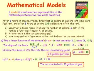
Associated assembly reactions Association constant: Factorise: : denotes the geometric degeneracy of the incoming subunit :denotes the degeneracy due to symmetry of the intermediates :denotes the bond energies R: gas constant T: temperature
Concentration profile One obtains the concentrations of the assembly intermediates via a recurrence relation:
Manipulating assembly behaviour: For different ratios a/c and b/c of the association constants, different assembly scenarios are obtained:
Summary and outlook We have developed new mathematical tools for the description of virus structure and assembly. • They are based on group theory and tiling theory, and have been used in order to model • the structure of viral capsids in terms tilings that encode the locations of the capsid proteins and the bonds between them • the structure of tubular malformations • the assembly process • the structure of the viral genome within the capsids • A symmetry principle that links different parts of the three-dimensional structure of viruses.
Outlook We are currently working on assembly models that include: • A 3d representation of proteins via encasing forms. • The role of RNA during assembly of RNA viruses. • Assembly via agglomeration of intermediates. • The dependence on experimental conditions (eg pH value) • Simultaneous assembly of different species. Applications include • the use of capsids for drug delivery. • interference with capsid assembly for anti-viral drug design. Financial support by an EPSRC Advanced Research Fellowship and EPSRC grant GR/T26979/01 are gratefully acknowledged.