Download
1 / 48
480 likes | 682 Vues
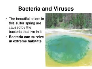
Microbial Models: The Genetics of Bacteria and Viruses. Chapter 18. Figure 1. Typical mosaic pattern on flue-cured tobacco leaves infected with Tobacco mosaic virus . (Courtesy H.D. Shew). Virus. LE 18-2. Bacterium. Animal cell. Animal cell nucleus. 0.25 µm. Capsomere of capsid.

E N D
Microbial Models: The Genetics of Bacteria and Viruses Chapter 18
Figure 1. Typical mosaic pattern on flue-cured tobacco leaves infected with Tobacco mosaic virus. (Courtesy H.D. Shew)
Virus LE 18-2 Bacterium Animal cell Animal cell nucleus 0.25 µm
Capsomere of capsid Membranous envelope RNA Capsomere Capsid LE 18-4 DNA Head RNA DNA Tail sheath Tail fiber Glycoprotein Glycoprotein 70–90 nm (diameter) 80–200 nm (diameter) 18 250 mm 80 225 nm 20 nm 50 nm 50 nm 50 nm Tobacco mosaic virus Adenoviruses Influenza viruses Bacteriophage T4
VIRUS Entry into cell and uncoating of DNA DNA Capsid LE 18-5 Transcription Replication HOST CELL Viral DNA mRNA Viral DNA Capsid proteins Self-assembly of new virus particles and their exit from cell Activity: Viral Reproductive Cycle
Lytic Cycle Attachment LE 18-6 Entry of phage DNA and degradation of host DNA Phage assembly Release Head Tail fibers Tails Synthesis of viral genomes and proteins Assembly Activity: Lytic Cycle
Phage DNA The phage attaches to a host cell and injects its DNA. Daughter cell with prophage LE 18-7 Many cell divisions produce a large population of bacteria infected with the prophage. Phage DNA circularizes Phage Bacterial chromosome Occasionally, a prophage exits the bacterial chromosome, initiating a lytic cycle. Lytic cycle Lysogenic cycle The bacterium reproduces normally, copying the prophage and transmitting it to daughter cells. Certain factors determine whether The cell lyses, releasing phages. Lytic cycle is induced Lysogenic cycle is entered or Prophage Phage DNA integrates into the bacterial chromosomes, becoming a prophage. New phage DNA and proteins are synthesized and assembled into phages. Activity: Lytic & Lysogenic Cycle
PRIONS = infectious proteins which cause degenerative brain diseases (like mad cow disease); most likely transmitted in food LE 18-13 Original prion Prion Many prions New prion Normal protein
Replication fork LE 18-14 Origin of replication Termination of replication
LE 18-17 Sex pilus 5 µm
Figure 18.15 Conjugation and recombination in E. coli (Layer 4)
Figure 18.17 Insertion of a transposon and creation of direct repeats
Insertion sequence LE 18-19 5¢ 3¢ 3¢ 5¢ Inverted repeat Inverted repeat Transposase gene Transposon Insertion sequence Insertion sequence Antibiotic resistance gene 5¢ 3¢ 3¢ 5¢ Transposase gene Inverted repeat
Regulation of enzyme production Regulation of enzyme activity Precursor Feedback inhibition LE 18-20 Enzyme 1 Gene 1 Gene 2 Enzyme 2 Regulation of gene expression Gene 3 Enzyme 3 Enzyme 4 Gene 4 Gene 5 Enzyme 5 Tryptophan
LE 18-21a trp operon Promoter Promoter Genes of operon DNA trpB trpA trpE trpC trpD trpR Operator Stop codon RNA polymerase Regulatory gene Start codon 3¢ mRNA 5¢ mRNA 5¢ D B E C A Inactive repressor Protein Polypeptides that make up enzymes for tryptophan synthesis Tryptophan absent, repressor inactive, operon on
DNA LE 18-21b_1 mRNA Active repressor Protein Tryptophan (corepressor) Tryptophan present, repressor active, operon off
DNA LE 18-21b_2 No RNA made mRNA Active repressor Protein Tryptophan (corepressor) Tryptophan present, repressor active, operon off
Promoter Regulatory gene LE 18-22a Operator lacl lacZ DNA No RNA made 3¢ mRNA RNA polymerase 5¢ Active repressor Protein Lactose absent, repressor active, operon off
LE 18-22b lac operon DNA lacl lacY lacA lacZ RNA polymerase 3¢ mRNA mRNA 5¢ 5¢ Transacetylase Permease -Galactosidase Protein Inactive repressor Allolactose (inducer) Lactose present, repressor inactive, operon on Activity: lac Operon
Promoter DNA lacl lacZ LE 18-23 RNA polymerase can bind and transcribe Operator CAP-binding site Active CAP cAMP Inactive lac repressor Inactive CAP Lactose present, glucose scarce (cAMP level high): abundant lac mRNA synthesized Promoter DNA lacl lacZ Operator CAP-binding site RNA polymerase can’t bind Inactive CAP Inactive lac repressor Lactose present, glucose present (cAMP level low): little lac mRNA synthesized
Capsid Capsid and viral genome enter cell RNA LE 18-8 HOST CELL Envelope (with glycoproteins) Viral genome (RNA) Template mRNA Capsid proteins ER Glyco- proteins Copy of genome (RNA) New virus
LE 18-11 The SARS-causing agent is a coronarvirus like this one (colorized TEM), so named for the “corona” of glyco-protein spikes protruding form the envelope. Young ballet students in Hong Kong wear face masks to protect themselves from the virus causing SARS.
Viral envelope Glycoprotein LE 18-9 Capsid RNA (two identical strands) Reverse transcriptase
Membrane of white blood cell HIV LE 18-10 HOST CELL Reverse transcription Viral RNA RNA-DNA hybrid 0.25 µm HIV entering a cell DNA NUCLEUS Provirus Chromosomal DNA RNA genome for the next viral generation mRNA Activity: HIV Reproductive Cycle New HIV leaving a cell