Download
1 / 86
900 likes | 1.42k Vues
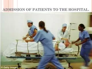
Approach to the Patients on Admission in Wards. Introduction of the New Internee. Aim:. To have clear conception of Medical emergency To have an idea of the referral system of the patients To be rational in the use of drugs To be rational in sending investigations

E N D
Aim: • To have clear conception of Medical emergency • To have an idea of the referral system of the patients • To be rational in the use of drugs • To be rational in sending investigations • To ensure better follow up of the patients
Thus, To provide better service to the patients
Admission During Office Hour Beyond Office Hour Through M.O.P.D Transferred/Referred Through Emergency Without NOD With NOD Brought dead Alive Psychiatric/ Functional Non psychiatric NOD= No Official Delay MOPD= Medicine out patient department
During Office Hour: • Patients come through MOPD, Outdoor and sometimes they are transferred from different wards. • Patients coming through emergency may have NOD note or may have admission ticket
In Case of NOD • Rush to the patient without any delay • Examine the patient, specially, vital signs (Pulse, Heart sound, Pupil, Planter reflex) to see whether the patient is alive or Dead • If you find the patient dead, show your sincerity to the attendants during examination though you are clinically certain that the patient is dead. • If found dead clinically, talk to the attendant/relative (specially with the 1st degree), start counseling. Tell them that your are almost sure about the death of the patient, and Now going to do an ECG just to confirm it!
5. Then do the ECG (This time, the Long Leads only) 6. If straight line is found, then address the EMO and declare the patient dead as Brought dead
If the patient is found gasping on the trolley, don’t waste time for the bed-head ticket to be available! Rather, immediately start the treatment with whatever resources you have. • Never forget to measure CBG of the Patients!
If CBG is found within normal range, you should be very much cautious regarding examination and diagnosis.
Because, Unconsciousness matters!!!!!
SIMULATED COMA • Psychologically disturbed patients sometimes feign coma. The eyes are actually closed and the patient is usually lying in a resting position, or supine with the arms and legs extended. • The eyelids resist attempts to open them • On forced eye opening, the eyes point upwards exposing the white conjunctiva (Bell's phenomenon) as part of the patient's attempt to maintain eyelid closure. • The eyelids close rapidly when released. The slow roving eye movements of organic coma cannot be simulated. • Painful stimuli to the limbs may be ignored, but pinprick to the nasal mucosa or to the lips usually elicits volitional grimacing. The pupillary light reflex is normal, as are plantar responses.
Cold caloric testing induces nystagmus with the fast phase away from the stimulated side, rather than deviation of the eyes toward the stimulus as would occur in true coma. Examination, especially invasive tests as above, may induce a return of cooperation and consciousness, or uncover a disturbed mental. (This is not practised in the ward)
In case of Functional disorder/Anxiety disorder/Acute stress disorder coming with NOD, Pseudo emergency may occur, like: Severe Chest Pain Psychogenic Hyperventilation, Respiratory distress Acute Mutism/ Unconsciousness Features: Typically can locate exactly with his/her own hand, particularly one finger! On pressing over the point, shout/cry out due to pain! X-ray and ECG (Tachycardia only) is normal Features: May complaints of Light headedness When excessive, tingling and numbness of limbs with carpopedal spasm may occur due hypocalcaemia resulting from Respiratory alkalosis X-Ray and ECG is normal Features: Typical eyeball movement with closure of eyelids! Few maneuver (NG tube insertion etc) may not be needed to treat them!
Just feel the pulse, hear the Heart sounds and look for pupillary reflex to make the attendants think that your are taking the case seriously • Then, address the EMO to admit the patient in the word • Meanwhile, send one of the attendants to bring some drugs (Just to make the attendants busy, other wise, they will make you Busy!) • Start counseling the attendants • Don’t talk/make any comment in front of the patient regarding your diagnosis (as it may cause exacerbation of symptoms)!
Management of the Patient: • In case of Functional disorder, I.V. drugs are given (or, sometimes need to be given) to make him/her think that treatment is started appropriately. • Drug list should include: • I.V. canula 20G • JMS infusion set • 1 inch Micropore • Inj. 5% DNS (If non diabetic) • Inj. Omeprazole • Inj. Dormicum/Inj. Haloperidol (suspecting that the patient may be restless or violent!)
Contd…….. • Sometimes, only oral medication is enough to treat the patient specially at night, when Staff nurses are few in number • Intravenous management is troublesome. • So, there is no reason to engage the sisters in treating those patient, rather we should think for really critically ill patients. • In that case, the medication will be: • Tab. Clonazepam (0.5mg ) /Tab. Midazolam 7.5 mg 1 Tab. stat and then 0+0+1 • Cap. Omeprazole (20mg) 1+0+1 [½ hr A/C] • Actually, it is the clinical condition and patient’s surrounding, which will lead you to the treatment.
Consciousnessis a state of normal cerebral activity in which the patient is aware of both self and environment and is able to respond to internal changes, for example hunger, and to changes in the external environment. • Altered consciousness resulting from brain disease may take the form of a confusional state, in which the patient's alertness is clouded; this is associated with \agitation, fright and confusion, i.e. disorientation. Such patients usually show evidence of misperception of their environment, and hallucinations and delusions may occur.
Confusional states must be carefully distinguished from aphasia, in which a specific disorder of language is the characteristic feature, and from continuous temporal lobe epilepsy, a form of focal status epilepticusin which the behavioural disorder is often accompanied by aphasia if the epileptic focus is left-sided. Usually this can be recognized by the occurrence of frequent but slight myoclonic jerks of facial and especially perioral muscles, and by variability in the patient's confusion from moment to moment during the examination. Always pause and observe an unconscious or drowsy patient for a few moments before disturbing them.
Abnormal drowsiness is often found in patients with space-occupying intracranial lesions or metabolic disorders before stupor or coma supervenes. The patient appears to be in normal sleep but cannot easily be wakened and, once awake, tends to fall asleep despite verbal stimulation or clinical examination. Further, while awake such patients can usually be shown to be disorientated. Higher intellectual function, such as the ability to perform abstract tasks or to make judgements, is disturbed. Stupor means a state of disturbed consciousness from which only vigorous external stimuli can produce arousal. Arousal from stupor is invariably both brief and incomplete.
Pupil: • Pupillary size and responsiveness to a very bright unfocused light beam (not the light of an ophthalmoscope) should be noted. If the pupils are unequal, a decision as to which is abnormal must be made. Usually the larger pupil indicates the presence of an oculomotor (third) nerve palsy, whether from damage to the oculomotor nerve by pressure and displacement or from a lesion in the mesencephalon itself. • Occasionally the smaller pupil may be the abnormal one, as in Horner's syndrome. If the larger pupil does not react to light it is likely that there is a partial oculomotor nerve palsy on that side. If the smaller pupil also fails to react to light this may be the midposition pupil of complete sympathetic and parasympathetic lesions, indicating extensive brainstem damage
In drug-induced coma and in most patients with metabolic coma the pupillary responses to light are normal. • Exceptions to this rule are glutethimide poisoning and very deep metabolic coma, in which the pupils may become dilated but only rarely become unreactive to light. In pontine and in thalamic haemorrhage the pupils may be very small (pinpoint pupils) and unreactive to light.
Bilateral pinpoint pupils occur with brainstem lesions, opiate and other drug intoxications, and with pontine infarction There is ptosis, dilatation of the pupil with absence of the light reaction, and slight lateral deviation of the eye.
There is ptosis and a small reactive pupil The eyes tend to 'look towards the tip of the nose' and the pupils are small; later they become large and unreactive as upper brainstem involvement follows
When brainstem death occurs the midbrain disturbance is manifest by midposition, fixed (unreactive) pupils with eye closure
PATTERN OF BREATHING • Alterations in the rhythm and pattern of breathing are an important aspect of the assessment of the unconscious patient.
CHEYNE-STOKES (PERIODIC) RESPIRATION • In Cheyne-Stokes respiration, breathing varies in regular cycles. A phase of gradually deepening respiration is followed, after a period of very deep rapid breaths, by a phase of slowly decreasing respiratory excursion and rate. Respiration gradually becomes quieter and may cease for several seconds before the cycle is repeated. Depressed but regular breathing at a normal rate occurs in most drug-induced comas, but Cheyne-Stokes respiration can occur in coma of any cause, especially if there is coincidental chronic pulmonary disease. Cheyne-Stokes breathing in a comatose patient is a sign of a large unilateral space-occupying lesion with brainstem distortion, for example subdural haematoma, or of bilateral lesions from other causes, for example cerebral infarction or meningitis.
KUSSMAUL RESPIRATION • Deep, rapid sighing breathing at a regular rate should immediately suggest metabolic acidosis. Metabolic or uraemia is the commonest cause of this acidotic (Kussmaul) breathing pattern, but a similar pattern may occur in some patients with respiratory failure, and in deep metabolic coma, especially hepatic coma.
CENTRAL PONTINE HYPERVENTILATION • Deep, regular breathing may also occur with rostral brainstem damage, whether due to reticular pontine infarction or to central brainstem dysfunction secondary to transtentorialherniation associated with an intra- or extracerebral space-occupying lesion. This breathing pattern is called central neurogenic (pontine) hyperventilation. Interspersed deep sighs or yawns may precede the development of this respiratory pattern.
Rapid shallow breathing occurs if central brainstem dysfunction extends more caudally to the lower pons. When medullary respiratory neurons are damaged, for example by progressive transtentorialherniation, irregular, slow, deep gasping respirations, sometimes associated with hiccups (ataxic respiration), may develop. In patients with raised intracranial pressure, this sequence of abnormal breathing patterns is often associated with other evidence of brainstem dysfunction, including a rising blood pressure, a slow pulse, flaccid limbs, absence of reflex ocular movements and dilatation of the pupils.
Changing patterns of respiration in an unconscious patient, particularly the development of central neurogenic hyperventilation, provide important and relatively objective evidence of deterioration. These changes in respiratory pattern may occur in structural lesions with raised intracranial pressure, in brainstem infarction, and less commonly in some varieties of metabolic coma, especially hepatic coma. They are indicative of progressive and potentially fatal brainstem dysfunction, but not of its causation.
Now, let us come back to the patient that has just entered into the ward!
Meanwhile, counseling should be done simultaneously regarding the prognosis of the patient. • Try to show pessimistic attitude to the attendant (Specially when you can understand that the patient is going to expire very soon) • After initial resuscitation, try to refer the case to the respective discipline (When indicated), e.g. CCU/ICU/Nephrology/Neuromedicine etc. (in the office hour only).
Medically unexplained somatic symptoms • Patients commonly present to doctors with somatic symptoms. Whilst these are often clearly associated with a medical condition, in other cases they are not. Symptoms may be disproportionate to, or occur in the absence of, a medical condition and are then often referred to as 'medically unexplained symptoms' (MUS). MUS are very common and occur in a quarter to a half of patients attending general medical outpatient clinics. Almost any symptom can be medically unexplained and common examples include: • pain (including back, chest, abdominal and headache) • fatigue • dizziness • fits, 'funny turns' and feelings of weakness.
Patients with MUS may receive a medical diagnosis of a so-called functional somatic syndrome, such as irritable bowel syndrome and may also merit a psychiatric diagnosis on the basis of the same symptoms. The most frequent psychiatric diagnoses associated with MUS are anxiety or depressive disorders. When these are absent, a diagnosis of somatoform disorder may be applied
Emergency (Non functional) Through Outdoor Transferred/Referred May involve various systems of the body alone, or simultaneously Cardiac Respiratory Gastro Endocrine Poisoning Infection Haematological CNS Renal Venomous snake bite OPC poisoning Others
Emergency patients commonly admitted in wards (Except poisoning) are: Cardiac Shock, LVF, MI, CHB, Hypertensive Crisis Respiratory Severe CAP, Acute severe asthma/COPD, Tension pneumothoraxResp. failure , Ex. of COPD, Cor-pulmonale CNS Encephalopathy, Stroke , Meningitis, Encehalitis, GBS with resp. distress/failure, Status Epilepticus etc. Gastro Severe acute Pancreatitis, EV rupture, Severe hemoptysis, Hypo. Shock, Perforation, Acute abdomen Endocrine DKA, Hypoglycemic attack, HONK, Addison’s crisis, Infection Septicaemia, shock, Severe malaria Haematological Septicaemia, shock, anemic heart failure ARF, Ureamic encephalopathy, LVF, Metabolic acidosis etc Renal
When patient comes through M.O.P.D • Usually these patients are admitted with some chronic disease, e.g. PUO and sometimes may be presented with acute exacerbation, e.g. Huge ascites in case of CLD, Constipation/vomiting in Ca-stomach etc. • Take proper history and fine out the causes of their admission this time, i.e. presenting complaints • Start thorough physical examination. • Share your findings to your colleagues
On Bed head Ticket • Fill up the front page with- (Necessary for disease profile and some Medico-legal condition) • Name of the patient: • Age: • Sex: • Address: • Provisional Diagnosis • Date and Time: • Doctor’s Signature (Preferably name)
On next page: • Presenting complaints: (Try to avoid mentioning more and irrelevant/non specific complaints) • 1. • 2. • 3. • History of present Illness: (Here, the modified salient feature should be written to save time) • Which should include: • Elaboration of positive findings • Mentioning of Important negative findings • Mentioning risk factors/co-morbid conditions
On physical examination:Try to mention the findings concisely, such as: • Appearance • Build • Anaemia • Jaundice • Cyanosis • Clubbing • Edema • Ascites • Dehydration • Pulse • BP • Temp • Heart • Lungs • GCS • Pupil • Planter reflex: • Deep jerks • Neck rigidity • Kernig’s sign • Engorged vein • Lymphadenopathy *Sometimes, additional findings should be noted in some particular diseases
Provisional diagnosis: • Try to be specific • Broad term can be used otherwise, e.g. Anaemia under evaluation, Acute febrile Illness, Acute Confusional State etc. • Never write the abbreviated form, like ACS, DVT, RA etc. • As soon as you are confirmed, try to mention the latest Diagnosis and omit the previous one. • Try to avoid confusing terms, e.g. Shock, Chest pain, Respiratory distress, Abdominal pain etc.
Before sending Investigations, seek their previous reports. • Always ask your senior colleague regarding investigations, because, it will reduce unnecessary wastage of time and money • Always ask the reason of sending the particular investigations to your respective senior colleague • Try to learn, in short, the basic pathogenesis of the disease, and go through the text later on. • Try to know the next plan of investigation • Treat the patient according to the diagnosis
Adv: • CBC • Urine R/M/E • CXR P/A view • Blood Urea • Serum Creatinine • Serum Electrolytes • USG of Whole Abdomen • Next Plan: • CT scan of Brain • CT guided FNAC • USG guided FNAC • Endoscopy of UGIT etc. • O/A on Date at Time:(Sample) • Diet: NBM/Liquid/Soft/Normal/Diabetic/Salt and fluid restricted/Protein restricted etc. • Bed rest in Propped up/Lateral/Semiprone position • O2 Inhalation 2L/min SOS • Inj. N/S 1ooo cc I.V. @ 1o d/min (If I.V. antibiotic to be given) • Inj. 5% DNS I.V. @10 d/min ( If not known to be diabetic and CBG is normal) • Oral/ Inj. Antibiotic……….. • Oral / Inj. Omeprazole (40mg) + 9cc D/W I.V. 12 hourly • Oral Anti pyretic [If febrile] • Oral anxiolytic (less potent) • Suppository antipyretic SOS [If fever > 102˚ For more] • Condom/Foley’s catheter [ In case of bed ridden patients] • Please monitor Daily I/O • Please monitor all vital signs regularly N Name of Doctor Date: ……..
If the patient is to be kept NBM If Parenteral Nutrition is to be given If NG tube feeding is to be given Fluids Others • [150 ml×2 hourly×10 feedings] • Special Milk; Dal; Soup; F. Juice; Dub water; etc Total 2500-3000 ml of fluid to be given If Diabetic If Non diabetic Vitamins and Electrolytes The rest of the fluid should be replaced by I.V Inj. 5% DNS 1000 cc+ Inj. Regular Insulin (U-100) or Other soluble Insulin 10 units I.V @ 25-30 drops/min Inj. 5% DNS 1000 cc I.V @ 25-30drops/min Inj. Vit B complex Inj. Vit C Extra electrolytes according to severity and deficiency Special condition deserves special fluids therapy
Half Neuralization in Diabetic patients: • InJ.5% DA/ Inj. 5 % DNS contains 5 gm of Glucose per 100 ml, so 1000 ml of those fluids contain 50 gm of glucose. • 1 U of soluble insulin can neutralize 2.5 gm of glucose • Therefore, full neutralization of 5% DNS 1000 ml requires (50÷2.5)= 20 U of insulin. • So, half neutralizaton requires 10 U of Insulin