BLEEDING DISORDERS
BLEEDING DISORDERS. HEMOSTASIS. 1. VASCULAR PHASE 2. PLATELET PHASE 3. COAGULATION PHASE 4. FIBRINOLYTIC PHASE. Hemostasis. BV Injury. Tissue Factor. Neural. Coagulation Cascade. Blood Vessel Constriction. Platelet Aggregation. Primary hemostatic plug. Reduced Blood flow.

BLEEDING DISORDERS
E N D
Presentation Transcript
HEMOSTASIS 1. VASCULAR PHASE 2. PLATELET PHASE 3. COAGULATION PHASE 4. FIBRINOLYTIC PHASE
Hemostasis BV Injury Tissue Factor Neural Coagulation Cascade Blood Vessel Constriction Platelet Aggregation Primary hemostatic plug Reduced Blood flow Platelet Activation Fibrin formation Stable Hemostatic Plug • Lab Tests • CBC-Plt • BT,(CT) • PT • PTT Plt Study Morphology Function Antibody
NORMAL CLOTTING Response to vessle injury 1. Vasoconstriction to reduce blood flow 2. Platelet plug formation (von willebrand factor binds damaged vessle and platelets) 3. Activation of clotting cascade with generation of fibrin clot formation 4. Fibrinlysis (clot breakdown)
CLOTTING CASCADE Normally the ingredients, called factors, act like a row of dominoes toppling against each other to create a chain reaction. If one of the factorsis missing this chain reaction cannot proceed.
VASCULAR PHASE WHEN A BLOOD VESSEL IS DAMAGED, VASOCONSTRICTION RESULTS.
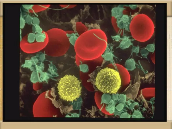
PLATELET PHASE PLATELETS ADHERE TO THE DAMAGED SURFACE AND FORM A TEMPORARY PLUG.
COAGULATION PHASE THROUGH TWO SEPARATE PATHWAYS THE CONVERSION OF FIBRINOGEN TO FIBRIN IS COMPLETE.
THE CLOTTING MECHANISM INTRINSIC EXTRINSIC Collagen Tissue Thromboplastin XII XI VII IX VIII X V FIBRINOGEN (I) PROTHROMBINTHROMBIN (III) (II) FIBRIN
FIBRINOLYTIC PHASE ANTICLOTTING MECHANISMS ARE ACTIVATED TO ALLOW CLOT DISINTEGRATION AND REPAIR OF THE DAMAGED VESSEL.
HEMOSTASIS DEPENDENT UPON: • Vessel Wall Integrity • Adequate Numbers of Platelets • Proper Functioning Platelets • Adequate Levels of Clotting Factors • Proper Function of Fibrinolytic Pathway
LABORATORY EVALUATION • PLATELET COUNT • BLEEDING TIME (BT) • PROTHROMBIN TIME (PT) • PARTIAL THROMBOPLASTIN TIME (PTT) • THROMBIN TIME (TT)
PLATELET COUNT • NORMAL 100,000 - 400,000CELLS/MM3 < 100,000Thrombocytopenia 50,000 - 100,000Mild Thrombocytopenia < 50,000Sev Thrombocytopenia
BLEEDING TIME • PROVIDES ASSESSMENT OF PLATELET COUNT AND FUNCTION NORMAL VALUE 2-8 MINUTES
PROTHROMBIN TIME • Measures Effectiveness of the Extrinsic Pathway • Mnemonic - PET • NORMAL VALUE • 10-15 SECS
PARTIAL THROMBOPLASTIN TIME • Measures Effectiveness of the Intrinsic Pathway • Mnemonic - PITT NORMAL VALUE 25-40 SECS
THROMBIN TIME • Time for Thrombin To Convert • Fibrinogen Fibrin • A Measure of Fibrinolytic Pathway NORMAL VALUE 9-13 SECS
So What Causes Bleeding Disorders? • VESSEL DEFECTS • PLATELET DISORDERS • FACTOR DEFICIENCIES • OTHER DISORDERS ? ?
VESSEL DEFECTS • VITAMIN C DEFICIENCY • BACTERIAL & VIRAL INFECTIONS • ACQUIRED & • HEREDITARY CONDITIONS
Vascular defect - cont. • Infectious and hypersensitivity vasculitides - Rickettsial and meningococcal infections - Henoch-Schonlein purpura (immune)
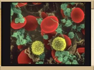
PLATELET DISORDERS THROMBOCYTOPENIA THROMBOCYTOPATHY
THROMBOCYTOPENIA INADEQUATE NUMBER OF PLATELETS
THROMBOCYTOPATHY ADEQUATE NUMBER BUT ABNORMAL FUNCTION
THROMBOCYTOPENIA • DRUG INDUCED • BONE MARROW FAILURE • HYPERSPLENISM • OTHER CAUSES
OTHERCAUSES • Lymphoma • HIV Virus • Idiopathic Thrombocytopenia Purpura (ITP)
THROMBOCYTOPATHY • UREMIA • INHERITED DISORDERS • MYELOPROLIFERATIVE DISORDERS • DRUG INDUCED
FACTOR DEFICIENCIES(CONGENITAL) • HEMOPHILIA A • HEMOPHILIA B • von WILLEBRAND’S DISEASE
FACTOR DEFICIENCIES • HEMOPHILIA A (Classic Hemophilia) • 80-85% of all Hemophiliacs • Deficiency of Factor VIII • Lab Results - Prolonged PTT • HEMOPHILIA B (Christmas Disease) • 10-15% of all Hemophiliacs • Deficiency of Factor IX • Lab Test - Prolonged PTT
FACTOR DEFICIENCIES • VON WILLEBRAND’S DISEASE • Deficiency of VWF & amount of Factor VIII • Lab Results - Prolonged BT, PTT
OTHER DISORDERS(ACQUIRED) • ORAL ANTICOAGULANTS • COUMARIN • HEPARIN • LIVER DISEASE • MALABSORPTION • BROAD-SPECTRUM ANTIBIOTICS
INHIBITORS 30% of people with haemophilia develop an antibody to the clotting factor they are receiving for treatment. These antibodies are known as inhibitors.These patients are treated with high does of FVIIa for bleeds or surgery. This overrides defect in FVIII or FIX deficiency. Longterm management involves attempting to eradicate inhibitors by administering high dose FVIII (or FIX) in a process called immune tolerance
Clinical Features of Bleeding Disorders Platelet Coagulation disorders factor disorders Site of bleeding Skin Deep in soft tissues Mucous membranes (joints, muscles) (epistaxis, gum, vaginal, GI tract) Petechiae Yes No Ecchymoses (“bruises”) Small, superficial Large, deep Hemarthrosis / muscle bleeding Extremely rare Common Bleeding after cuts & scratches Yes No Bleeding after surgery or trauma Immediate, Delayed (1-2 days), usually mild often severe
Platelet Coagulation Petechiae, Purpura Hematoma, Joint bl.
Petechiae (typical of platelet disorders) Do not blanch with pressure (cf. angiomas)Not palpable (cf. vasculitis)
Petechiae in patient with Rocky Mountain Spotted Fever
Ecchymoses (typical of coagulation factor disorders)
CT scan showing large hematoma of right psoas muscle
Inheritedbleedingdisorders Hemophilia A and B vonWillebrands disease Other factor deficiencies Acquiredbleedingdisorders Liver disease Vitamin K deficiency/warfarin overdose DIC Coagulation factor disorders
Hemophilia A and B Hemophilia A Hemophilia B Coagulation factor deficiency Factor VIII Factor IX Inheritance X-linked X-linked recessive recessive Incidence 1/10,000 males 1/50,000 males Severity Related to factor level <1% - Severe - spontaneous bleeding 1-5% - Moderate - bleeding with mild injury 5-25% - Mild - bleeding with surgery or trauma Complications Soft tissue bleeding
Hemophilia Clinical manifestations (hemophilia A & B are indistinguishable) Hemarthrosis (most common) Fixed joints Soft tissue hematomas (e.g., muscle) Muscle atrophy Shortened tendons Other sites of bleeding Urinary tract CNS, neck (may be life-threatening) Prolonged bleeding after surgery or dental extractions
Treatment of hemophilia A • Intermediate purity plasma products • Virucidally treated • May contain von Willebrand factor • High purity (monoclonal) plasma products • Virucidally treated • No functional von Willebrand factor • Recombinant factor VIII • Virus free/No apparent risk • No functional von Willebrand factor