Septic Shock Management
Septic Shock Management. EBL Group 1 Carol, Marina, Sophia & Wendy.


Septic Shock Management
E N D
Presentation Transcript
Septic ShockManagement EBL Group 1 Carol, Marina, Sophia & Wendy
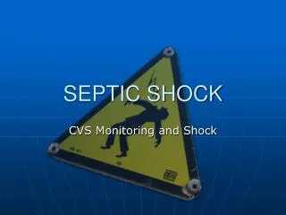
Patients with severe sepsis (which can progress to septic shock) develop an imbalance between oxygen delivery and demand that results in global tissue hypoxia. If this is not corrected, the result is multi-organ failure and death.
Treatment of Septic Shock • Provide oxygen, and relieve respiratory distress • Give fluids by IV to restore blood volume • Give vasoactive drugs to treat low blood pressure • Treat underlying infections with antibiotics and surgery (if needed) • Support any poorly functioning organs • Reverse abnormal blood clotting with drugs
Treatment during the first hour after diagnosis of Septic Shock • Baseline observations of vital signs including heart rate, BP, temperature, respiratory rate, SpO2 and level of consciousness. • Blood specimens for FBC, U&E, glucose and lactate, clotting screen, blood cultures and LFTs. • Arterial blood gas measurements • Blood cultures should be taken before antibiotics are given from a peripheral vein and where possible using IV lines already in place (e.g. Hickman line)
Lactate, an important indicator of tissue perfusion, should be measured even if there are no signs of hypotension. Bicarbonate (HCO3) concentration of less than 20mmol/L seen on ABG or U&E result is a warning sign that lactate concentration is high. • IV broad spectrum antibiotics should be administered promptly. • IV fluid is administered if systolic BP is less than 90mmHg or mean arterial pressure (MAP) less than 65mmHg. A fluid challenge (500ml given over 5-10 minutes is repeated up to a total of 2 litres)
If the patient remains hypotensive, insertion of a central venous line and continuation of IV fluid challenges to achieve a CVP of between 8 and 12mmHg should be considered. If after receiving between 1.5 and 2 litres of fluid, the patient remains hypotensive with a MAP of less than 65mmHg, an IV infusion of the vasoconstrictor, noradrenaline should be administered and an arterial line inserted to monitor BP accurately.
A urinary catheter that allows hourly measurements of urine output should be inserted and a specimen of urine obtained. • The source of sepsis should be found.
The patient’s temperature is usually elevated in the early stages of septic shock and they may commonly experience shaking chills. As the shock progresses, the temperature typically drops and the patient experiences diaphoresis (profuse perspiration).
Haemodynamic monitoring • The evaluation of the pressures in the heart and lungs may be required.
Central Venous Catheters • Inserted into one of the central veins (superior or inferior vena cava). These two veins return blood into the right atrium and have the largest blood flow in the body. • Insertion sites - Internal Jugular, subclavian vein, femoral vein or brachial vein - this is usually used for peripherally inserted catheters (PICC).
Central Venous Pressure • Central Venous Pressure (CVP) is the filling pressure of blood returning to or filling the right atrium. Venous pressure reflects venous return to the heart. CVP is the pressure in the superior or inferior vena cava as the blood enters the heart and indicates blood volume, vascular tone and cardiac function.
Complications of insertion • Arrhythmias - this occurs during insertion especially when the introducer guide wire makes contact with the tricuspid valve or the catheter is inserted too far. • Pneumohaemothorax - can result from accidental puncture of the pleura during the procedure. The patient’s haemodynamic and respiratory status must be monitored closely. • Haematoma - caused by trauma to the vein and or surrounding tissues. An artery may be accidentally punctured by the introducer.
Catheter in wrong position - occasionally the catheter may follow a path from the heart towards the head or down the arm. This will not give an accurate CVP reading. • Air Embolus - this may occur if the catheter is not properly connected to the appropriate tubing or if one of the portals or 3 way taps is left open to air. • Central vein thrombosis - occurs in 4 to 35% of patients who have a central line (as a result of good or poor practice). Therefore, clinical areas have accepted the introduction of heparin coated lines to reduce thrombus formation (Wright et al 2001).
Measuring the CVP • The CVP may be measured using a transducer system or manually using a manometer (uncommon in ICU as it is far less accurate). • A tranduced catheter will give continuous display of the CVP on a monitor. The wave form should show small undulations reflecting the changes in pressure within the right atrium during the cardiac cycle. • Normal CVP pressure is 3-10 mmHg.
Steps for measuring a patient’s CVP • Ensure all IV infusions running through the manometer line are stopped. • Zero the manometer. • Close the 3 way tap to the patient, opening the tap between the fluid and the manometer. • Allow the manometer to fill with fluid to a level beyond the expected pressure. Do not fill the line so far that the air filter becomes wet. • Close the tap to the fluid source opening the manometer to the patient. • Watch the fluid level change. It should fall until gravity pressure equals the pressure from the central veins. When the fluid stops falling read the CVP measurement.
Conditions affecting the CVP • Increased values: • Right ventricular failure • Pericardial tamponade • Fluid overload • Pulmonary hypertension • Tricuspid regurgitation • Pulmonary stenosis • Peripheral vasoconstriction
Decreased values: • Hypovolaemia • Peripheral vasodilation (including Sepsis)
Central Venous Catheters should be changed within 7 days, if infection is suspected a different site should be used with a new catheter. • If the Central Venous Catheter site looks inflamed or infected a swab should be sent for microbiology culture and sensitivity.
Arterial Catheters • This is an invasive device with potential complications. • The arterial catheter can facilitate the high level care needed by critically ill patients. • Can be used for patients who require continuous monitoring. • The readings and waveforms are dynamic and change with every beat of the cardiac cycle (BP monitoring). • Also used to obtain arterial blood samples (ABG). • This also provides Mean Arterial Pressure (MAP) essential to approximate the perfusion of essential organs such as the kidneys. • This also records arterial pressure waveform (APW).
Arterial catheter insertion sites: • Radial artery • Femoral artery • Brachial artery • Dorsalis pedis • Inserted by medical staff or anesthetist. Nurses would assist with this procedure.
Complications • Haemorrhage • Thrombosis • Infection • Haemorrhage is the most dangerous complication which can result in large blood loss in a short space of time. It is important to label the line ‘Arterial’ to prevent confusion and the misuse of the line e.g. administering drugs.
As well as managing numerous advanced therapeutic interventions, nursing priorities of care include strict attention to the use of universal precautions and prevention of secondary infection.
Nursing Management • Close monitoring of the patient using MEWS • Awareness of normal/abnormal observation results • Ensure emergency equipment is available and checked • Psychological care (support for patient and relatives) • Information • Management of skin integrity • Nutritional intake • Personal care / mouth care • Patient positioning
Scary !!!!!!!!! • If prompt recognition and management of symptoms is not instigated, the patient may progress to the irreversible stage of shock and organ damage does not respond to treatment. • Therefore prompt recognition, assessment and management of septic shock optimizes outcome for the patient.
References • Adam, K. Osbourne, S. (2005) Critical Care Nursing Science and Practice. 2nd edition. Oxford: Oxford University Press. • Bench, S. (2004) Clinical Skills: assessing and treating shock: a nursing perspective. British Journal of Nursing. 13 (12) p.715-721. • Critical Care Nursing made incredibly easy (2004) London: Lippincott, Williams and Wilkins. • Garretson, S. (2005) Haemodynamic monitoring: arterial catheters. Nursing Standard. 19 (31) p.55-64. • Medline (2006) Septic Shock. [online] available from: http://www.nlm.nih.gov/medlineplus/ency/article/000668.htm [accessed 2 Feb 2006].
Robson, W. Newell, J. Beavis, S (2005) Severe sepsis in A&E. Emergency Nurse. 13 (5) p.24-27. • Robson, W. Newell, J. (2005) Assessing, treating and managing patients with sepsis. Nursing Standard. 19 (50) p.56-64 • Woodrow, P (2002) Central venous catheters and central venous pressure. Nursing Standard. 16 (26) p.45-51. • Woodrow, P. (2004) Arterial Blood Gas Analysis. Nursing Standard. 18 (21) p. 45-52. • Wright, F. et al (2001) Antibiotic coated central lines: do they work in the critical care setting? Clinical Intensive Care. 12 (1) p.21-28.