Download

1 / 42
610 likes | 1.44k Vues
MUCOCUTANEOUS INVOLVEMENT IN Systemic Lupus Erythematosus. By: Samah Hamdy El- Medany Ass. Lecturer in Rheumatology & Physical medicine and Rehabilitation dep. . INTRODUCTION.

E N D
MUCOCUTANEOUS INVOLVEMENT IN Systemic Lupus Erythematosus By: SamahHamdy El-Medany Ass. Lecturer in Rheumatology & Physical medicine and Rehabilitation dep.
INTRODUCTION • Lupus Erythematosus (LE) is a difficult disease to classify in terms of skin findings because it can cause many different types of skin lesions, and the challenge is to determine how these lesions come about, how they fit together, and how best to treat them. By understanding the basis of these skin lesions, scientists can develop more effective therapies.
INTRODUCTION Gilliam (1977) initially proposed a classification system for the skin lesions that can be encountered in patients with LE and divided the cutaneous manifestations of this disease into those that are histologically specific for LE (i.e., LE-specific skin disease) and those that are not histologically specific for this disease (i.e.,LE-non-specific skin disease). In 2003, a modified classification system of cutaneous LE (CLE) has been developed including acute CLE, subacute CLE, chronic CLE, and the intermittent subtype of CLE.
Classification of Lupus Erythematosus–Associated Skin Lesions Modified from Sontheimer RD, Provost TT: Cutaneous Manifestations of Rheumatic Diseases. Baltimore, Williams & Wilkins, 1996.
CLASSIFICATION OF LUPUS ERYTHEMATOSUS–ASSOCIATED SKIN LESIONS Modified from Sontheimer RD, Provost TT: Cutaneous Manifestations of Rheumatic Diseases. Baltimore, Williams & Wilkins, 1996.
Classification of Lupus Erythematosus–Associated Skin Lesions Modified from Sontheimer RD, Provost TT: Cutaneous Manifestations of Rheumatic Diseases. Baltimore, Williams & Wilkins, 1996.
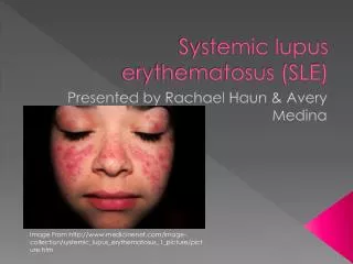
Photosensitivity refers to the development of a rash after exposure to ultraviolet B (UVB) radiation found in sunlight or fluorescent lights. It occurs in 60% to 100% of patients with SLE. The severity of cutaneous reaction depends on the intensity of the UV source and the duration of exposure. This image displays the cheeks and nose of a patient affected by systemic lupus that has been aggravated by exposure to the sun. Photosensitivity The dull red patches of systemic lupus are prominent in sun-exposed areas. When these patches are seen on the cheeks, one can imagine why the term “butterfly rash” is used. This image displays how inflammation in systemic lupus can be intense, causing very red skin lesions.
Pathophysiology of photosensitivity in LE Lupus erythematosus (LE) represents an autoimmune disease with great clinical variability in which photosensitivity is a common feature for all forms and subsets. Cutaneous LE lesions often arise in sun-exposed areas and it is well reported and recognized that sun exposure may also exacerbate or induce systemic manifestations of this disease.
Amplification cycle demonstrating UV-injury, apoptosis, necrosis, and chemokine production. Mediation of recruitment and activation of autoimmune T-cells and INF-alfa producing Plasmocytoiddendritic cells (PDCs). Release of more effector cytokines amplifying chemokine production and leukocyte recruitment leading to LE-lesion. (From Meller 2005).
Acute cutaneous lupus erythematosus • Acute cutaneous lupus erythematosus (ACLE) usually occurs in association with systemic manifestations preceding by weeks or months the onset of a multisystem disease. • Sun exposure is a common exogenous factor to precipitate ACLE. Furthermore, infections, especially with subtle types of viruses, or certain drugs, e.g. hydralazine, isoniazide, and procainamide, have also been found to induce or aggravate this disease.
Acute Cutaneous Malar Rash Note Sparing of Nasolabial Folds Acute cutaneous lupus erythematosus • There are localized and generalized manifestations of ACLE. The localized form commonly presents as the classic ‘‘malar rash’’ or ‘‘butterfly rash’’ on the central portion of the face and may only affect the skin transiently. Therefore, at the onset of disease, the patient may mistake this rash for sunburn. It usually begins with small, discrete erythematous macules and papules, occasionally associated with fine scales and gradually becomes confluent and hyperkeratotic. Facial swelling may be severe in some patients; however, it mostly disappears without scarring and pigmentation.
Acute cutaneous lupus erythematosus • Similar lesions have also been found to occur on the forehead, the V-area of the neck, the upper limbs, and the trunk. In addition, patients may have diffused thinning or a receding frontal hairline with broken hair (lupus hair), and may further present with teleangiectasias and erythema of the proximal nail fold. Superficial ulcerations of the oral and/or nasal mucosa are also frequently accompanied with this subtype and may cause extreme discomfort in some patients.
Acute cutaneous lupus erythematosus • The generalized form of ACLE is a less common variety and may be located anywhere on the body although the preferred sites are above the waistline. The onset of this form usually with exacerbation of systemic manifestations developing a prolonged disease activity. • The incidence of this generalized form is estimated to be approximately in 5–10% of patients with SLE. • It is characterized by a symmetrically distributed maculopapular or exanthematous eruption with a pruritic component. The colour of the lesions is usually red or, less frequently, dull red or livid, and there have been reports of patients presenting with severe involvement of the oral mucosa or the palms and phalanges
SUBACUTE CUTANEOUS LUPUSERYTHEMATOSUS Subacute cutaneous lupus erythematosus (SCLE) is not uniformly associated with SLE. About 50% of affected patients have SLE, and about 10% of patients with SLE have this type of skin lesion. Patients with SCLE may present with annular or psoriasiform skin lesions, and this is strongly associated with anti-Ro (SS-A) and anti-La (SS-B) antibodies. Patients with SCLE have a high incidence of photosensitivity and rarely can present with erythema multiforme–like lesions (Rowell's syndrome). Most patients with SCLE have prominent cutaneous and musculoskeletal complaints but generally do not develop a severe systemic disease.
Subacute cutaneous lupus lesions. Typical features include symmetric, widespread, superficial, and non-scarring lesions. Involvement of the neck, shoulders, upper chest, upper back, and extensor surface of the hand is common. These lesions begin as small photosensitive, erythematous, scaly papules or plaques that evolve into a papulosquamous (psoriasiform) or annular polycyclic form as in this patient.
Subacute cutaneous lupus erythematosus (SCLE).Annular, polycyclic lesions with erythematous borders and central hypopigmentation on the back.
Another example of subacute cutaneous lupus erythematosus with annular lesions.
Chronic cutaneous lupus erythematosus 1- Discoid lupus erythematosus • Discoid lupus erythematosus (DLE) is the most common subtype of the chronic cutaneous variants of LE and may present as a localized or disseminated form. • The localized form, characterized by limited cutaneous involvement of the head and scalp, usually accounts for 70% of patients with DLE, and the disseminated form, characterized by the extension to areas below the neck for 30% of patients with DLE. • About 30% of patients with SLE may develop DLE lesions during the course of their disease and, in about 5–10% of patients, DLE lesions may already be present at the onset of the disease.
Discoid lesions are characterized by discrete, erythematous, slightly infiltrated plaques covered by a well-formed adherent scale that extends into dilated hair follicles (follicular plugging). Discoid lesions are most often seen on the sun-exposed parts, face, neck, scalp, ears and infrequently on the upper torso. They tend to expand slowly with active inflammation at the periphery, and then to heal, leaving depressed central scars, atrophy, telangiectasias, and dyspigmentation (hyperpigmentation or hypopigmentation). Chronic cutaneous lupus erythematosus Discoid LE
Discoid LE Chronic CLE Facial discoid lupus erythematosus lesions produce large areas of disfigurement on confluence. Note the erythema (indicating disease activity), keratin-plugged follicles, and dermal atrophy. The characteristic pattern of hyperpigmentation at the active border and hypopigmentation at the inactive center. Facial involvement of this sort can produce extreme psychosocial disability.
(a) (b) (c) Discoid LE Discoid lupus erythematosus on the finger (a), palm (b), and beard area (c).
Discoid LE Examples of DLE of the scalp with characteristic plugging of follicles, and demonstrating marked scarring and pigment change. This situation is irreversible.
Discoid LE Chronic cutaneous lupus erythematosus • About 2% of patients with DLE show a hyperkeratotic type of lesion consisting of dull, red, and indurated lesions. • When the palms and soles are involved, the mobility can be difficult. • Mucous membrane involvement can be found in 25% of patients with DLE, but does not necessarily reflect systemic manifestation or high disease activity. • The differential diagnosis of discoid lesions includes hypertrophic lichen planus, eczema, and actinic keratosis; some early and scaly discoid lesions also must be differentiated from psoriasis.
Discoid LE Chronic cutaneous lupus erythematosus Biopsy specimens of skin lesions from patients with DLE contain immune complexes at the dermal-epidermal junction. The basilar epithelium in these areas is vacuolated and edematous, and the dermis contains an inflammatory infiltrate.
Chronic cutaneous lupus erythematosus 2- Chilblain lupus erythematosus (CCLE) • The pathogenesis of this rare subtype is unknown, but microvascular injury secondary to exposure to cold and possible hyperviscosity from immunologic abnormalities may play a role. • The risk of developing SLE is estimated to be approximately 20%. • The lesions of this type are clinically characterized by symmetrically distributed, circumscribed painful areas of livid and purple plaques.
Chronic cutaneous lupus erythematosus 2- Chilblain lupus erythematosus • Mostly, the dorsal and lateral parts of the hands, feet, ears, nose, elbows, knees, and calves are involved.
Chronic cutaneous lupus erythematosus 3- Lupus erythematosuspanniculitis (profundus) • lupus profundus, presenting as a firm nodular lesion with or without an overlying cutaneous lesion. The nodules are often painful and consist of perivascular infiltrates of mononuclear cells plus panniculitis, manifested as hyaline fat necrosis with mononuclear cell infiltration and lymphocytic vasculitis. The nodules usually appear on the scalp, face, arms, chest, back, thighs, and buttocks; ulcerations are uncommon, and they usually resolve leaving a depressed area. Some patients with lupus profundus show no other manifestations of SLE. Multiple, erythematous nodules and indurated plaques were present on the lower extremities. On the right thigh and medial left knee there were skin-colored, atrophic, scarred plaques.
Lupus erythematosuspanniculitis (profundus) An older lesion of lupus profundus, demonstrating depression of the skin surface due to marked atrophy of the underlying fat.
INTERMITTENT CUTANEOUS LUPUS ERYTHEMATOSUS Lupus erythematosustumidus (LET) • Tumid lupus, a rare variant, is characterized by photodistributed lesions with chronic pink-to-violaceous papules, nonscarring plaques, and nodules. • Tumid lupus differs from other variants of CLE. Scarring which is the hallmark of DLE, does not occur in tumid lupus. Hypopigmentation, frequently evident in patients with SCLE with erythema and scaling, has never been detected in tumid lupus A round erythematous edematous plaque on the right malar region
Single. erythematous, succulent, urticaria-like plaques on the forehead Intermittent cutaneous lupus erythematosus LET The prognosis in patients with LET is generally more favorable than in those with other forms of CLE
Involvement of the mucous membranes occurs in 25% to 45% of patients with SLE. The most common manifestations include irregularly shaped, raised, white plaques; areas of erythema; silvery white scarred lesions; and ulcers with surrounding erythema on the soft or hard palate or buccal mucosa. The oral ulcers in SLE are usually painless, and sometimes there is no apparent association between their presence and systemic disease activity. Oral lesions may be the first signs of SLE. Mucosal LE
Characteristic discoid lesions with erythema, atrophy, and depigmentation can occur on the lips. Nasal ulcers have been noted in patients with SLE. They usually are found in the lower nasal septum, tend to be bilateral, and are associated with active disease. Nasal septum perforation has been reported in 4% of SLE patients and is secondary to vasculitis. Involvement of the upper airway mucosa also can occur and cause hoarseness. Mucosal LE
Alopecia (nonscarring) Papulonodular mucinosis Livedo reticularis Vasculitic lesions NON - SPECIFIC CUTANEOUS MANIFESTATIONS OF LUPUS ERYTHEMATOSUS
Erythema multiforme Dermatitis herpetiformis Pemphigus erythematosus Raynaud's phenomenon Skin lesions which are seen not only in patients with LE but are also found in association with other conditions are defined as non-specific cutaneous manifestations. Non – Specific CLE
TYPES OF LUPUS ERYTHEMATOSUS AND THEIR DIFFERENTIAL DIAGNOSIS
If a skin rash is present, the doctor may take a biopsy (a tissue sample) from the margin of a skin lesion. A test known as a lupus band detects antibodies known as immunoglobulin G (IgG), which are located just below the outer layer of the tissue sample. They are present in about 80% of patients with active SLE and in between 30 - 40% of those with inactive disease. The biopsy will not differentiate between systemic and discoid lupus, but it can rule out other diseases. Skin Tests Microscopic image of direct immuno-fluorescence using a fluorescent anti-IgG antibody on a skin biopsy. The test shows a band-like accumulation of IgG along the basement membrane ("lupus band test positive“)
TREATMENT OF CUTANEOUS LUPUS ERYTHEMATOSUS • Sunscreens are important therapeutically in all types of LE, especially SCLE and tumid LE. • The usual treatment for discoid lesions is a potent topical corticosteroid. • Intra-lesional steroid injections may be useful for refractory discoid lesions, or isolated areas of LE profundus. • Antimalarials, usually hydroxychloroquine, are a useful therapy in many patients with SLE (especially if photosensitivity is prominent), and especially in SCLE.
PRACTICE POINTS • Always consider discoid lupus erythematosus (DLE) in any patient with scarring alopecia and evidence of inflammation (perifollicular erythema at the margin of scarred areas). • A butterfly rash without malaise or other systemic symptoms is unlikely to represent active systemic lupus erythematosus (SLE). • If you suspect SLE in a patient with photosensitivity and DLE-like lesions, but the ANA test is negative, check for anti-Ro antibodies: the patient may have (SCLE). • Patients on antimalarials for lupus erythematosus must stop smoking to get good benefit.
Conclusions • Cutaneous manifestations in patients with LE are very frequent, show a great variety and can occur at any stage of the disease. • A classification system has been established dividing the skin lesions associated with LE in specific and non-specific manifestations. • Patients who have a more generalized involvement of the skin tend to have more systemic symptoms than those with lesions localized to the face or neck. • All cutaneous manifestations of LE can result in limited patient quality of life and disability
REFERENCES • Kelley's Textbook of Rheumatology, 8th ed. (2008) • The Skin in Systemic Autoimmune Diseases (2006) • http://www.lupus.org/webmodules/webarticlesnet/templates/new_aboutintroduction.aspx?articleid=75&zoneid=9 • http://en.wikipedia.org/wiki/Systemic_lupus_erythematosus • http://www.lupusmalaysia.org/e/what-is-sle/ • http://www.cureresearch.com/l/lupus/stats-country.htm