Download
1 / 32
520 likes | 1.8k Vues
Systemic Lupus Erythematosus (SLE). Clinical features A chronic autoimmune disease with variable tissue injury in multiple organs, including kidney, brain, skin, joints, heart, lungs, muscles and blood

E N D
Systemic Lupus Erythematosus (SLE) • Clinical features • A chronic autoimmune disease with variable tissue injury in multiple organs, including kidney, brain, skin, joints, heart, lungs, muscles and blood • Strong genetic predisposition MHC and non MHC immune response genes, females>> males (>10/1) • The onset may be insidious or fulminant, typically appearing in a previously healthy person in adolescence or young adulthood • The course is characterized by multiple flares and remissions • Therapy involves intensive use of high dose corticosteroids and alkylating agents or other non specific immunosuppressivedrugs
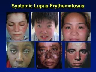
Major Clinical Manifestation of SLE ManifestationPercent Arthralgias/arthritis 95 Hematological 90 Rash 81 Fever 77 Neurologic 59 Renal 53 Pulmonary 48 Cardiac 38
Lupus Erythematosus: Epidemiologic findings • Primarily a disease of young adults Peak incidence age 15-45 • Marked female preponderance Sex ratio during peak incidence is 10 : 1, female : male • Distinctive ethnic distribution Greatly increased incidence among African-Americans (0.3%), compared to Caucasoids or Blacks in Africa. Similarly increased incidence among Hispanics, Mestizo Indians in Mexico, Sioux Indians, and generally among Chinese and Filipinos, but not among most other Asian peoples.
Systemic Lupus Erythematosus (SLE) • SLE is the prototypic systemic autoimmune disease where the dominant autoimmune response is the production of an array of autoantibodies to self antigens including nuclear components (DNA, RNA, histones) as well as autoantibodies to cell membrane determinants on hematopoietic cells including (RBC, platelets and leukocytes). • The autoantibodies induce injury by forming immune complexes with autoantigens which deposit in vessels walls to cause vasculitis and glomerulonephritis. The auto antibodies may also directly bind to cell membranes and destroy cells by activating complement killing and by triggering FcR mediated inflammatory and cytotoxic mechanisms. • The B cell autoantibody response is in turn driven by MHC-restricted CD4+ T cells that recognize self peptides likely bound by HLA-DR2 & DR3 MHC molecules.
Clinical features of SLE which reflect an ongoing immune response • • lymphadenopathy with active germinal center formation, splenomegaly • • polyclonal hypergammaglobulinemia • • anti-nuclear antibodies, anti-dsDNA, multiple antibodies to other nuclear structures • lymphopenia, thrombocytopenia, hemolytic anemia • cytokine mediated systemic phenomena: fever , malaise, weight loss (TNFa, IL-1b)
Autoantibodies in SLE • I. Antibodies to DNA • double-stranded DNA (unique to SLE) • double- and single-stranded DNA • single-stranded DNA • II. Antibodies to Deoxyribonucleoprotein • Antigen: complex of DNA and histone • III. Antibodies to Other Nuclear and Cytoplasmic Constituents • histones • nonhistone nuclear proteins • a. Small nuclear ribonucleoproteins(snRNPs) • Sm antigen (SLE specific), Ro / La, • b. ENA (not specific for SLE), RNA • IV. Antibodies to Cell Membrane Antigens • red blood cells, platelets • T cells, B cells, macrophages, granulocytes • b2 microglobulin, cardiolipin • V.. Antibodies to soluble proteins • Anti-Antibodies: rheumatoid factors • Anti-b glycoprotein 1, clotting factors
Antinuclear autoantibodies in SLE Anti dsDNA antibodies are highly specific for SLE Rim ANA pattern on Hep 2 cells that accompanies anti dsDNA antibodies, may include anti lamin and anti Ku Anti dsDNA staining of Crithidia kinetoplast. Very specific
Antinuclear autoantibodies in SLE Anti histone and anti DNA antibodies (nucleosome) Anti ds DNA Anti histone (>50-75%), Specific for SLE (30-40%)SLE, not specific for SLE, >90% in drug induced lupus Anti ssDNA, non specific
Many SLE autoantigens are large complexes of RNA and multiple proteins Anti Ro 52kD, Ro 60kD, La • Four small uridine-rich RNA molecules, hY1, 3, 4, 5 RNA variably associate with Ro and La proteins Anti Ro 52kD and anti 60kD 60% SLE, 90% Sjogren’s syndrome Anti La 15% SLE, 60% Sjogren’s syndrome
Infarction of distal vessels by reactive vascular proliferation and occlusion induced by deposition of immune complexes • May occur in nearly any organ
Lupus Nephritis Diffuse, segmental proliferative / necrotizing glomerulitis, Class IV >50% of glomeruli involved with endocapillary or mesangial hypercellularity, epithelial crescents, or fibrinoid necrosis Large subendothelial deposition of immune complexes in glomerular basement membrane Clinically: Most severe. Renal insufficiency in >50%. Red cell casts, hematuria and HBP.
CD4+ T cell Driving force Autoreactive to self peptides Autoreactive B cells IgG autoantibody Production Autoantibody-mediated Disease SLE pathogenesis Environmental triggers (drugs, microbes ?) Genetic susceptibility: Complex polygenic Genes Involved: MHC class II Complement deficiency Multiple non-MHC (unknown) X-chromosomal (unknown) Self-antigen driven Other genetic influences ?
Lupus Erythematosus Strong familial aggregation: 25% cases have affected blood relative. 50% concordance of identical twins Genetic associations MHC genes: HLA-DR2, and HLA-DR3 (DRB1*0301) MHC genes: C2, C4(?) deficiency Polymorphism of FcRIIa and FcRIII Fas gene deficiency
Two major mechanisms of antibody-mediated tissue injury operating in SLE
Clinical features attributable to SLE autoantibodies reacting with cell surface structures or soluble proteins
Disease: Vasculitis Glomerulitis Arthritis Pleuritis Pericarditis Dermatitis Serum Sickness develops after injection of soluble foreign antigens
Nephritis, vasculitis Nephritis, vasculitis Anti dsDNA autoantibodies CH50 SLE course reflecting presence of immune complex disease
An important normal function of complement is to regulate the disposition of immune complexes • C1q binds to IgG in complex and activates C3 • C3b attaches and mediates binding of the complex to CR1 (CD35) on red blood cells • The immune complexes are solubilized or transported to the spleen on RBC where the immune complexes are phagocytosed and degraded by macrophages and removed from the circulation
If excess immune complexes are not physiologically cleared they deposit in tissues and initiate inflammatory programs • Interact with FcR or CR on circulating or tissue cells (Monocytes, macrophages, neutrophils, NK cells, etc.) and initiate a receptor mediated proinflammatory program, e.g. leukocyte mediated killing, cytokine release & vasculopathy • Deposit in blood vessel wall or in glomerulus where initiate inflammation by either interacting with complement and CR of a tissue cell, or interacting directly with FcR on the tissue cells, initiating a receptor mediated proinflammatory program resulting in immune complex disease
Several genetic diseases emphasize the importance of a normal complement system in preventing autoimmunity • Inherited C1q deficiency strongly predisposes to SLE, perhaps through a central role of C1q in handling disposal of apoptotic cells • Inherited C2 deficiency results in a disease with many features of SLE, but without nephritis • The MHC haplotype HLA-A1-B8- DR3 strongly predisposes to SLE. This haplotype contains a defective C4 gene in the class III region of the MHC as well as the known HLA-DR3 susceptibility gene
The role of FcR- immune complex interactions in mediating inflammation and immune injury in SLE • • Immune complexes interact with Fc receptors to initiate a receptor mediated proinflammatory program • Polymorphism of FcRIIa and FcRIII in humans affect affinity of these FcR for IgG and influence the occurrence and severity of nephritis in SLE • FcRIII a or g-chain plays a critical role in initiating immune complex inflammation in mice with spontaneous autoimmune diseases as shown by the absence of this pathway of injury in FcRIII “knockout” mouse strains despite the presence of immune complexes
Two similar mechanisms of immune complex glomerulitis Deposition of preformed soluble complexes In situ formation of complex on “planted” autoantigen
Why do SLE patients make autoantibodies? (1) Anti-self immunity: abrogation of self tolerance SLE might be the result of insufficient elimination of autoreactive T cell clones in the thymus or periphery. This might result in such autoreactive T cells being released into the peripheral circulation and causing the autoimmune features of the disease (2) Hidden antigens The nuclear and cytoplasmic antigens that are associated with autoimmunity are not commonly exposed to the immune system. If such antigens (dsDNA, for example) are liberated during cellular turnover, they may incite an immune response. Thereafter, further release of such antigens might form the nidus for IC
Why do SLE patients make autoantibodies? (3) Cross reactivity SLE might be a disease caused by an unknown pathogen such as a virus or a bacterium. The interaction of pathogen derived peptides with a susceptible HLA haplotype may elicit "autoimmune" diseases by activating pathogenic T cells. Such a pathogen has not been identified in SLE, but no feature of the disease suggests that this could not be the etiology. (4) Abnormal regulation: failure of suppression SLE might arise as a consequence of abnormalities in regulatory CD4+ or CD8+ T cells.
Evidence that T cells are important in the development of SLE • The pathogenic anti-DNA antibodies in SLE are high affinity IgG molecules. Because it is known that class switching to IgG as well as somatic mutation and affinity maturation requires T cells we infer that anti-DNA antibody-producing B cells are expanded in SLE by a process that mimics the normal CD4+ T cell-dependent responses, involving common mechanisms of somatic mutation, affinity maturation, and IgM to IgG class switching. • The MHC class II restriction and the known association of DR2 and DR3 with susceptibility to SLE also strongly point to a predominant role CD4+ T cells in the induction of autoimmunity in SLE. • Finally, animal models of SLE are effectively treated with molecules which block key functions of CD4+ T cells.
Antigen Presentation by B cells in SLE SmIg Anti-DNA DNA Antigen MHC class II carrier protein (histone) MHC class II/ histone peptide complex B cell histone peptides Antigen binds specifically to SmIg, is internalized into vesicles and cleaved into peptides which displace and bind to MHC class II molecules. The peptide/MHC complex is then transported to the surface membrane.
Expression of Membrane Proteins Following Antigen Specific Activation of T and B Cells SmIg TCR Resting T cell Resting B cell MHC class II CD4 IL-2 CD 23 SmIg IL-2R TCR Activated T cell CD 40 CD40L CD4 MHC class II CD80 CD86 CD28 Activated B cell MHC class II lymphokines
CONSEQUENCES OF CD40L/CD40 INTERACTIONS DURING T-B CELL INTERACTIONS CD23 CD40L TCR Sm Ig CD4 CD40 Activated T cell ActivatedB Cell • Triggering of B cell proliferation • Rescue from apoptosis • Induction of Ig isotype class switching • Up-regulation of B71 and B72 • Germinal center formation • Up-regulation of CD23 • Downregulation of CD40L expression
CD23 SmIg CD40 Activated B cell IgG IgA IgE Plasma Cell Final Phases of B cell Differentiation are Mediated by Contact T cell signals (CD40L/CD40) and Lymphokines CD40L TCR CD4 Activated T cell Lymphokines IL-2, IL-4, IL-5, IL-6, IFN-g, TGFb
Molecular Interactions of Helper T Cells and APC/B Cells: Potential targets of therapy for SLE CD4+ T Cell CTLA-4 CD28 CD3 p56 lck CD2 z z g d e h h CD40L TCR C C LFA-1 a b CD45 V b V a peptide B7 LFA-3 B7 CD4 MHC II ICAM-1 APC/ B cell CD20 CD40