More upper extremity
More upper extremity. IMAGE REVIEW & CRITQUE Lecture # 3 – Part 1 Upper Extremity RT 123 – WEEK 9. Shield ?. Anatomy &Positioning Review. Upper Limb. RE: LATERALS PG 80 PROJECTION VS POSITION. CHEST / ABDOMEN / SKULL


More upper extremity
E N D
Presentation Transcript
More upper extremity IMAGE REVIEW & CRITQUE Lecture # 3 – Part 1 Upper Extremity RT 123 – WEEK 9
Anatomy &Positioning Review Upper Limb
RE: LATERALS PG 80PROJECTION VS POSITION • CHEST / ABDOMEN / SKULL • The lateral is referred to the POSTION – which is the SIDE TOUCHING THE IR • SO LEFT LATERAL (position ) in referred to as a LATERAL PROJECTION • LIMBS : Pos/Pro – side entered by IR • MEDIOLATERAL OR LATERALMEDIAL
LEFT LATERAL OF CHEST LEFT LATERAL POSITION – LATERAL PROJECTION
Lateromedial humerus Mediolateral humerus Taken “AP” “Taken PA”
EPI’s ? Hand Position? Hand - lateralmedial Hand - mediolateral Internal Rotation - (lesser tubercle in profile)
Greater tubercle Lesser tuberclein profile inexternal rotation internal rotation Proximal HUMERUS
? Position – Best Seen?
supracondylar fracture of the elbow Name 3 postions to best demonstrate this fx?
supracondylar fracture of the elbow If a child complains of elbow pain after a fall and refuses to straighten his or her arm
Patient positioning for AP humerus image when fracture is located close to shoulder.
Lateromedial humerus Mediolateral humerus Taken “AP” “Taken PA”
Distal Humerus (poss fx)Poor position distally - better position not good Rad Prot
It is useful to have some idea of the age at which each of the carpal ossific centres appears although one would not expect you to know them all by heart! Ossification is usually visible by the end of the first year in the capitate and hamate . The remainder of the carpals, except for the pisiform, have appeared by the eighth year. Ossification of carpals
The capitate (1) and hamate (2) both are seen as large circular bony structures. A very faint smaller bony ossific centre is also present just proximal to the hamate. This is the first appearence of the triquetral The epiphyseal growth plates have also started to ossify on each of the metacarpals. These are at the proximal ends of these bones. Bone age?
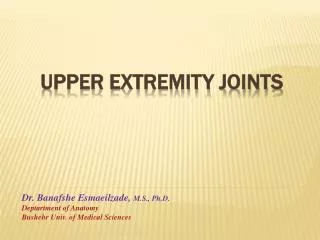
GLIDING BALL & SOCKET HINGE SADDLE JOINT CLASSIFICATION
fracture description • The first consideration is the age of the patient. • Fractures in children require special attention because a child's bones are still growing and changing. • An adult fracture is classified based on its location, direction, alignment, articular involvement (involving most of the joint rather than the shaft), and whether it is open or closed.
The direction of the fracture is described as • transverse (fracture line is straight across the bone) • spiral (fracture line spirals down the bone) • oblique (slanted fracture line) • comminuted (more than two fragments) • segmental (several large fractures in the same bone) • open fracture means that bone fragments have broken through the skin causing an open wound • closed fracture means that there is no opening in the skin.
Fracture Terminology Review • Greenstick fractures – occurs when bone is angulated beyond limit of bending • Complete fractures - Transverse fractures • - Spiral / Oblique Comminuted / Segmental • Avulsions: • Forcible tearing/separation of ligaments or muscles from the bone
COMMON FRACTURES PG 99 • Avulsion – tearing away from bone • Boxer – base 4th or 5th metacarpal • Colle’s – distal radius/ulnar – posteriorly • Smith’s distal radius/ulnar – anteriorly • Greenstick – bending of bone (children) • Pathologic – fx of a diseased or weakened bone • + Galeazzi and Monteggia fractures
Upper Extremity Avulsions • Avulsion of muscles • Greater tubercle • Lesser tubercle/ • Medial epicondyle of humerus • Lateral epicondyle of humerus
Epiphyseal plate fractures: 30% of children fractures involve the growth plate (epiphyseal plate Children
Buckle (torus) fractures – caused by compression failure of bones. It occurs usually near the metapysis where porosity is greatest
The most difficult aspect of reconstruction of fractures of the distal humerus is the restoration of normal anatomic relationships
MORE IMAGE ANALYSISPractice TEST “FILM CRITIQUE” WHAT DO YOU KNOW?