Download

1 / 22
260 likes | 698 Vues
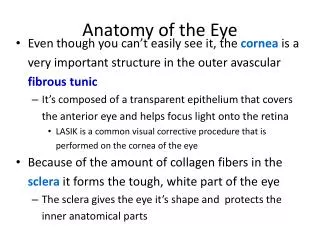
Anatomy of the Eye. California State Society for Opticians. Anatomy of the Eye. A fun approach to learn about the eye is to draw. The following slides are examples from webpage sources to practice drawing. Example drawing of the Eye. Practice fill-in. Index of Refraction of the Eye

E N D
Anatomy of the Eye California State Society for Opticians
Anatomy of the Eye • A fun approach to learn about the eye is to draw. • The following slides are examples from webpage sources to practice drawing.
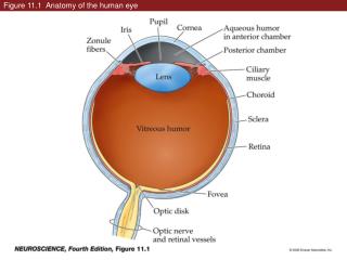
Index of Refraction of the Eye Cornea – 1.37 Crystalline Lens – 1.42 Aqueous Humor – 1.33 Vitreous Humor – 1.33
Diopter of the Eye Cornea – 43 Crystalline Lens – 17 Total Diopter Power: 60
Note where the Cones & Rods are located above. Cones (How Many) Day Vision Detail Color Rods: (How Many) Night Vision Peripheral Vision Black/White
Write out the Layers of the Cornea Listed on slides 8, 9 & 10 – information from webpage sources. It is important to memorize the layers of the cornea.
CORNEA BASICSThe cornea is approximately .5 mm thick and contains two-thirds of the eye’s total focusing power. As light enters the eye, it is refracted (bent) by the cornea and the lens until it refocuses on the retina. The convex surface of the cornea is thicker in the periphery than in the center, and the size remains static after the first year of life. Since the cornea does not contain a blood supply, it obtains oxygen from the tears that coat the eye. The tears of the eye obtain oxygen from air which is distributed throughout the tear film. The oxygen assists in maintaining overall corneal health by keeping the surface clear and shiny. Even though there are no blood vessels, the cornea is very sensitive due to the numerous nerve endings extending into it.For many years, the cornea was known to contain five layers; however there has been a recent discovery of a sixth layer. What follows is a description and discussion of the various layers as well as their functions. EPITHELIUM The epithelium is a five-to-seven-level-thick structure that assists in maintaining corneal optical clarity by constant regeneration. It is composed of flattened cells that are replaced continuously and provide a smooth surface for the tear film. The tear film helps provide nutrients to the front surface of the cornea and washes out foreign objects from the eye. Another function of the epithelium is to block foreign material from entering the area.There are thousands of nerve endings in the epithelium that cause the cornea to be extremely sensitive to pain. It is bathed by the tears of the eye, which keep the cornea moist. Other functions of the tear film are to maintain the smooth surface of the epithelium, assist in optical clarity, and provide nutrition to the cornea. Injury to the corneal epithelium causes sloughing off of the epithelial cells resulting in corneal abrasion. Since these cells regenerate quickly, the cornea is able to heal itself rapidly. Deeper penetration into the cornea by an injury can lead to scars which can cause the cornea to lose its optical clarity and visual acuity.
BOWMAN’S LAYER The very tough condensed layer of cells that lie below the epithelium of the cornea is called the Bowman layer. It is composed of collagen fibers that are difficult to penetrate. As a result, this layer aids in protecting the cornea from deeper injury. An injury to this area of the cornea results in scarring, which if located in the visual axis can affect acuity.STROMA Lying directly beneath Bowman’s layer is the stroma. This is the thickest part of the cornea and is composed of 16% collagen and 78% water. The collagen in this layer run parallel to one another and is very strong and elastic. The fibers here do not contain blood vessels. They are evenly spaced and contribute in maintaining corneal transparency as well as the cornea’s shape.DUA’S LAYER Located just below the stroma is the newly discovered corneal layer called Dua’s layer. This tier was discovered in May 2013 by HarminderDua, MD, professor of ophthalmology and visual sciences at the University of Nottingham. The layer was discovered by Dr. Dua’s team while doing research on corneal transplantation on donated eyes. Dua’s layer is very thin—15 microns thick—and is impervious to air which Dr. Dua and his colleagues believe may play a major role in patients undergoing corneal transplant surgery. Perforation and tears to this layer can cause fluid buildup in the cornea and result in corneal hydrops. Therefore, further research in the study of this layer can be useful in the diagnosis of patients with keratoconus.
DESCEMET’S MEMBRANE The thin, very elastic fifth layer of the cornea is Descemet’s membrane which acts as a protective barrier against infection. It is comprised of very elastic collagen fibers that are different from those in the stroma which retract if cut. It also serves as a protective barrier against infection and injury to the eye. This membrane is 8–10 microns in thickness, is extremely resilient, and will regenerate if damaged. This membrane is located beneath the stroma and supports the endothelium and is important to the health of the endothelium. ENDOTHELIUM The sixth and most posterior layer of the cornea is the endothelium. This level lies adjacent to the anterior chamber and is the innermost layer of the cornea. This single layer of cells maintains the balance of water in the cornea by keeping it in a partially dehydrated state, by pumping water from the stroma. This fluid balance is essential for the clarity of the cornea.
Eye Muscles - 6 Memorize the 6-eye Muscles
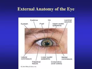
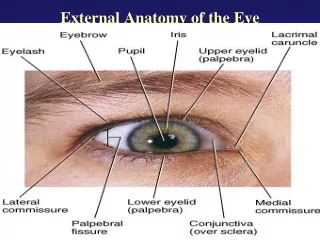
Structure & Function the Eyes • The Sclera (sk-LAIR-uh) is the white of the eye. It provides a skeletal framework of tough, leather-like connective tissue that the retina, choroid, and extraocular muscles attach to. • The Anterior Chamber – space between the posterior cornea and the anterior iris that is filled with aqueous humor. • The anterior chamber angle is located at the point where the iris and cornea intersect. This is the area where aqueous humor filters/drains out of the eye to maintain even levels of intraocular pressure (IOP)
Structure & Function the Eyes • Posterior Chamber – the space immediately behind the iris, extending to the front of the crystalline lens. This space is filled with a fluid known as aqueous humor. • Aqueous (ay-KWEE-us) humor helps to nourish the cornea & the lens, as well as maintaining the pressure inside the eye. • Crystalline Lens (KRIS-tah-lin) – flexible transparent structure just behind the pupil • Second refracting organ that helps to direct light rays to focus on the retina. • Flexible and changes shape (and power) depending on the distance from the object viewed to the eye. • Long & flat when looking at distant object, short & thick when focused on near object
Structure & Function the Eyes • Iris – colored part of the eye. Regulates the amount of light allowed into the eye – has 2 rings of muscle fibers that allows it to regulate light entering the eye • Dilates (enlarges)pupillary opening to allow more light in under dark conditions. This is known as MYDRIASIS (iris dilator muscle does this) • Constricts (shrinks) pupillary opening to reduce the amount of light into the eye in bright conditions. This is known as MIOSIS (iris sphincter (sf-INC-ter) muscle does this) • One of the 3 structures that makes up the uveaIris+ciliarybody+choroid= uvea
Structure & Function the Eyes • Vitreous (VIT-ree-us) Chamber - This space is filled with a clear gel-like substance that helps to give the globe its shape. Fills the posterior 2/3 of the eyeball • Retina (RET-ih-nuh) - Specialized light-sensing tissue where light comes into focus. Similar to film in a camera. It transfers this focused light to the optic nerve. • Macula Lutea (MAC-yoo-luh loo-TEE-uh) - Vital specialized area of the retina that is responsible for sharp central vision
Structure & Function the Eyes • Choroid (KOR-oyd) - Vascular tissue made of blood vessels that nourishes the retina. One of the 3 parts that make up the uvea (U-vee-uh). Iris+ciliarybody+choroid= uvea • Optic Nerve – contains over 1 million nerve fibers • Converts light energy into electrical signals that are carried in the optic nerve to the brain. • One of the 12 cranial nerves. We mainly deal with 6 of the 12 cranial nerves