Pediatric Hematology & Oncology: Understanding Anemia in Children
690 likes | 735 Vues
Learn about different types of anemia, their causes, symptoms, and treatments in children, explained by Dr. Isam Haddadin, a pediatric hematologist and oncologist based in Amman, Jordan.

Pediatric Hematology & Oncology: Understanding Anemia in Children
E N D
Presentation Transcript
Anemias Dr. Isam Haddadin Pediatric hematologist / Oncologist Amman-Jordan
Anemia is defined as a reduction of the red cell volume or hemoglobin conc. Below the range of the values for age occurring in healthy person.
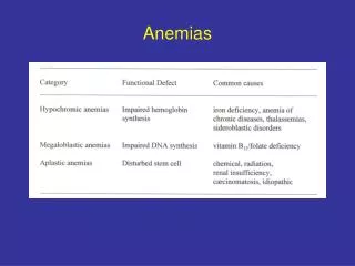
Classification of Anemias • Inadeguate intake • Production( Bone Marrow) • Hemolytic Anemias • Blood Loss
Inadequate intake • Deficiency of specific factors a. Iron def. b.Vit. B12 c. Folic acid
Production 1. Congenital pure red cell Anemia 2. Anemia of infection a. Renal failure b.Cancer 3. Ineffective Erythropiosis Cong.D.A
Hemolytic Anemias RBC defects : Spherocytosis Elliptocytosis Enzyme defects : G6PD pyruvate kinase Defect in synthesis of Hb : HbS , C , D , … Thalassemia Immunologic : Rh incompatibility ABO incompatibility
Physiology of Anemia in infancy • Newborn : higher Hb • Physiologic anemia : 2-3 months • Premature baby : exaggerated anemia
Megaloblastic Anemia Presence of Megaloblasts in BM and Macrocytes in the blood. Folic acid def : Inadequate intake,poverty,Goats milk Absorption-congenital or acquired Increase Requirement;Hemolysis Disorder of Metabolism;MTHFR def. Onset : 4-7 months (infant)
Megaloblastic Anemia Vit. B12 def. : -Inadeguate intake;Diet or mother -Defective absorption,Intrinsc factor -G.I.T;malabsorpion,surg,crohn disease -Defective B12 transport;Cong TCII def -Acquired; drugs Age : 9 month – 10 years Lab : Macrocytic , pancytopenia,hypersegmented neutrophil,Megaloblastic BM
Clinical Manifestation • Failure to thrive • Generalized Weakness • Glossitis • Anorexia • Pallor • Jaundice • Neurolgic Manif;Ataxia,paresthesia,hyporeflexia
Treatment of Megaloblastic Anemia 1. Folic acid : 5 mg Tab/day Higher dose in cong. def. 2. Vit. B12 : I mg I.M daily for 2 weeks (if neurologic defect,subacute dorsolateral degeneration of cord),then monthly. 3.Vit.B12: 200-1000 microgm/week x 4 Then monthly 4.Transcobalamin II def;Large doses Vit.12
Iron def. Anemia Most common 5.5% among chlid Requirement;2 mg/kg/day .Peak at 9-24 months • Inadeguate intake;Diet,Breast milk • Impaired absorption;Celiac • Blood loss ; G.I;Mickel,cow milk allergy,Liver,Lung
Iron Deficiency Anemia 525x Microcytic, Hypochromic Anisocytosis polychromasia
Tissue Effect Of Iron Def. Anemia • 1.G.I: Anorexia,pica,atrophic glossitis,dysphagia,Guaiac positive • 2.CNS: Irritability,fatique decrease activity,breath holding spells. • 3.Cardiovascular: Tachycardia,cardiac hypertrophy and H.F • 4.Immne system: Increase infection,impaired granulocytes killing.
Diagnosis OF Iron Def. • History and physical examination;Diet,Family hist,Pallor, No hepatosplenomegaly • CBC; Hb,PCV,MCV,MCH,MCHC are low and RDW is >15. • Platelets are increased. • Blood Film:Hypoch,Microcytic,Anisocytosis,poikiloct • Serum Ferritin<12 ng/ml. Iron level is low • Diff Diag: Thalassemia,Lead poisoning,Sideroblstic anemia,Chronic infection,Malignancy
Treatment Of Iron Def • 1.Diet;fortified with iron milk,Cereals,Vit,C, • 2.Rx:Oral Ferrous; Dose 6.omg/kg/day.3 doses and betwee meals.Duration for 2-3 month. • 3.Response: Increase appetite,then Retic count,Hb and the stores. • 4.Failure to Response: poor compliance Blood loss Inadequate dose Coexistent disease,malabsorption, thyroid,malignancy,VitB12 def. 5.Parenteral Rx: Iron Dextran I.M in Bowel disease , no compliance
Hemolytic Anemia : Evidence of Hemolysis • PCV • Bilirubin indirect • Retic count • haptoglobin
Hereditary Spherocytosis • Aut- Dominant: 75% , Mutation : 25% • Defect in cell membrane : spectrin ,Ankyrin • Clinical: Mild HS,Moderate Anemia ,Severe HS 10% Splenomegally,Gall stones . • Lab: Low PCV,MCV High MCHC,RDW,Retic.Spherocytosis Osmotic Fragility test positive . • Present at newborn as Hemolytic Anemia, Jaundice • Management ; Blood transfusion, splenectomy
G6PD Glucose-6-phosphate Dehydroginase Deficiency • X-linked , more in male • Most common RBC enzyme defect • Hemolysis with oxidant stress( infection,drugs,fava beans) • Drugs ; Asprin , Sulfa , Nitrofurantion, • Hb precipitation,RBC membrane damage leading to premature red cell destruction. .Mediterranean type Severe. Others like US usually moderate. .Lab: Evidence of hemolysis .Management:Obtain sample,Remove Oxidant,Transfusion
Heinz Bodies • Heinz bodies are denatured globin, and represent the end-product of oxidative degradation of haemoglobin. • Presence of stained inclusions close to the red cell membrane (Heinz bodies). supravital stain
Immune Hemolytic Anemia Extracorpuscular H.A • Isoimmune; Rh-Incomp. And ABO- Incomp. • .Autoimmune: Idiopathic-----IgG warm Ab IgM cold Ab. Secondary-----Infection;EBV,CMV Drugs---Cephalosp,Penicillin Malignancy---Lymphoma • Lab:CBC, Retic , Coombs test,Spherocytes. • Treatment: Steroid Transfusion IVIG Splenectomy Immunotherapy . .
Hematological Manifestation Of Systemic Diseases • Results from: • 1.BM dysfunction—Anemia,Polycythemia • Thrombocytopenia,Leukopenia,Leukocytosis • 2.Hemolysis • 3.Immune Reaction • 4.Altered in Hemostasis, Inhibitors to factors
Hematologic Manif. Cont… 1 .Heart: Prosthetic valves—Thrombocytopenia Anemia due to hemolysis, Iron def. 2.G.I.T: Reflux, Stomach(Vit.B12),Celiec, Bowel disease, Bleeding(Mickles) 3Liver : Hypersplenism,Wilson disease Shortened RBC survival,Bleeding. 4.Renal: Erythropoeitin,Uremia,Blood loss Dialysis,RBC survival decreased. 5.Chronic Illness: Decrease Fe flow from RES. 6.Skin: DyskeratosisCongenita,Eczema
Thalassemia Introduction: Beta thalassemia major affects 60000 births per year worldwide. A mutation in the B-globin gene causes defective erythopoiesis ,which in its homozygous form results in a severe anemia ,rendering the individual dependent on lifelong blood transfusions .
Alpha Thalassemia 4-genes (~ 600 mutations) Beta Thalassemia 2-genes (25 mutations) (~80% of mutations) Major Classification of Thalassemia
If both parents are Thalassemia Carriers 25% chance of contracting thalassemia major 50% chance of contracting thalassemia minor 25% chance of being Normal Chances to have Thalassemia
Thalassemia Major Clinical • -Chronic hemolytic anemia • -Skeletal deformaties • -Splenomegaly • -Heart failure • -Iron overload • -Gall stones • -Growth impaired
B-Thalassemia • .Lab:1. Microcytosis,Hypochromia.Anemia , Target cell. • 2.Hb-electrophoresis ; HbF increased Hb A1 decrease Hb A2 normal • Normal values: Hb A1 95% HbA2 2-3.5% Hb F 1%
B-Thalassemia • Treatment: • 1.Chronic Blood Transfusion. • 2.Folic Acid. • 3.Chelation Therapy. • 4.Splenectomy. • 5.BMT.
Prevention of Thalassemia 1. Identification of carriers (long Term prevention) 2. Genetic counselling of families with known Thalassemia “Clinical”for couples at risk before marriage(practical term of prevention) 3. CBC for Couples; If MCV less than 78 ,do Hb-electroph. to role out Thal. Trait.
Prevention of Thalassemia Prenatal Screening Of Couples at Risk after Conception (not practical)
Carrier IdentificationStandard scheme 1. Peripheral blood count and indices: (R.B.C, Hb , MCV, MCH, MCHC, RDW) done by automated counter 2. Hb-electrophoresis
ProcedurePrenatal diagnosis 1. CVS* < 8-12 weeks of gestation. 2. AFT** > 15 weeks of gestation. • Study fetal cells for parent’s same mutation of thalassemia minor they have * : Chorionic villus sampling ** : Amniotic fluid testing
-Pakistan: 5.5% (4-7 million estimated carriers) -Jordan: 4.5% Gaza strip: 3.5% -Iran: 3.64%(8000 pregnancies at risk/year) -Iraq:3 million carriers, ? 25000 cases. -Sri-lanka : 2.0%, 1000 cases thalassemia major -Italy : 8000 cases of thalassemia major Incidence of Thalassemia world wide (Carrier’s)
Sickle Cell Anemia .Aut.recessive,Valine substituted for Glutamic acid in the No.6 position of B chain. .Sickle cell Trait has benign clinical course .8 % American blacks have the trait. .HbS about 40%.Severe hypoxia may produce Vaso-occlusive phenomena. .Carriers should avoid hypoxia.
Sickle Cell Anemia Sickle Cell Disease: Homozygous for the gene .Presentation until one year of age. Clinical Manifestation: 1.Painful Vaso-occlusive crises; Hands,Feets 2.Strokes due to cerebral occlusion. 3.Acute Chest Syndrome. 4.Sequestration crises, pooling of blood in spleen and liver—State of shock. 5.Aplastic crises ,Parvovirus. 6.Sepsis; capsulated organism
Sickle CELL • Lab: Hb 5-9gm/dl. Spontaneous sickling, Target cells Retic 5-15%,Bil increased, Howell Jolly bodies Sickling test . Hb-electroph; HbS 90 % Hb F 2-10% Hb A 0% Hb A2 2.5%
Sickle Cell Anemia Sickle cells Boat Cells Target Cell
Sickle-Thalassemia Peripheral blood: S-thalassemia- In Sickle-thalassemia, the anemia tends to be milder than in SS disease, there are few sickle cells, and there is microcytosis. 640x
Sickle cell Treatment 1.Therapy during episodes 2.Tonics: Folic acid.e 3.Analgesics: Paracetamole, Codeine,Morphine 4.Antibiotics; Penicillin, Vaccin 5.Hydration during crises 6.Blood Transfusion not always,ExchangeTransfusin in Acute chest syndrome 7.Splenectomy in hypersplenism or sequestration. 8.Hydroxyurea. 9.BMT; in certain cases
Hypertransfusion Protocol : Hb level 10.5 gm (pretransfusion level) 1.Maximizing growth and development 2.Minimizing extramedullary hematopoisis and decreasing skeletal abnormalities 3.Reduce iron absorption from gut 4.Reduce splenomegaly and hypersplenism
Iron overload 1.Ongoing transfusion therapy 2.Increase gut absorption of iron 3.Chronic hemolysis
Chelation Therapy The objectives are : 1.To bind free extracellular iron 2.To remove excess intracellular iron 3.To attain a negative iron balance (i.e, iron excretion > iron input)
Iron Chelating Agents Desferal 1.Iron binding efficiency 1:1 2.Iron slectivity is high 3.Regimen Sc or iv infusion 4.Dose 40mg/kg 5.Safety: good, poor compliance
Iron Chelating Agents Ferriprox 1.Binding 3:1 2.Iron selectivity some report of zinc def. 3.Regimen : oral, three times daily 4.Safety neutropenia, joint problems 5.Dose 75mg/kg
Iron Chelating Agents ICL670(Ex jade) 1.Iron binding 2:1 2.Iron selectivity highly selective 3.Regimen oral, once daily 4.Safety skin rashes, kidney toxicity 5.Dose 20mg/kg