QUIPPS II
50 likes | 65 Vues
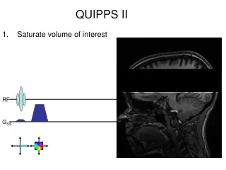
This technique involves saturating the volume of interest, tagging incoming blood, killing the bottom half of the bolus, and waiting for the rest to enter the volume of interest. The image read-out follows this pulse. The time it takes for the tagged bolus to reach the volume of interest is recorded. The perfusion image is obtained by subtracting the control image from the tagged image, with the inverted spins from labeled blood decreasing net magnetization.

QUIPPS II
E N D
Presentation Transcript
QUIPPS II • Saturate volume of interest RF GSS
QUIPPS II • Saturate volume of interest • Tag (invert) incoming blood RF GSS
QUIPPS II • Saturate volume of interest • Tag incoming blood • Kill bottom half of bolus RF GSS
QUIPPS II • Saturate volume of interest • Tag incoming blood • Kill bottom half of bolus • Wait for the rest to enter VOI • Image Read-out follows this pulse. Time it takes tagged bolus to reach VOI RF GSS
QUIPPS II • Perfusion image = control minus tagged • In tagged image, inverted spins from labeled blood decrease net magnetization