Spinal Control of Movement
110 likes | 268 Vues
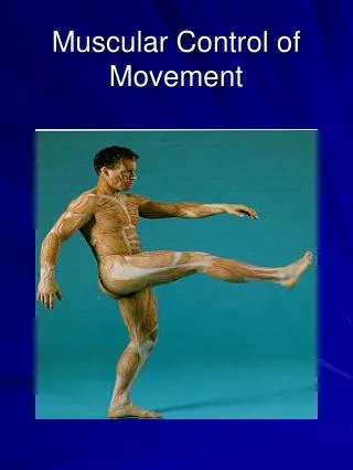
Spinal Control of Movement. Midterm 2 Results. Types of Muscles. Smooth – digestive tract, arteries Striated: Cardiac – accelerates or slows heart rate Skeletal – moves bones around joints, moves eyes, facial expression, respiration, speech

Spinal Control of Movement
E N D
Presentation Transcript
Types of Muscles • Smooth – digestive tract, arteries • Striated: • Cardiac– accelerates or slows heart rate • Skeletal – moves bones around joints, moves eyes, facial expression, respiration, speech • Skeletal muscles are the somatic motor system and are under voluntary control.
Types of Movement • Extension – takes limb away from body (opens penknife) • Flexion – brings limb toward body (closes penknife) • Muscles cannot push so any movement requires coordination • Synergists – muscles that work together • Antagonists – muscles that pull in opposite directions • Muscles are also named by location: • Axial (trunk), proximal (shoulder, elbow, knee), distal (toes)
Motor Units • Each muscle fiber is innervated by an alpha motor neuron. • Bundles of fibers form large and small motor units. • Small motor units act first and control fine motor movement. • Finer control is possible under lighter loads. • Fast contracting, fast fatiguing white fibers form “fast” motor units (slow ones are red). • Alpha neuron firing rate makes a fiber/motor unit fast or slow.
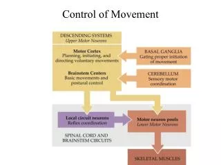
Input to Alpha Motor Neurons • Dorsal root ganglion input to muscle spindle. • Provides feedback about muscle length. • Upper motor neurons in the motor cortex and brain stem. • Voluntary control of muscles. • Interneurons
Muscle Fiber Structure • Muscle fibers are enclosed by an excitable cell membrane called sarcolemma. • Within the muscle fiber are cylindrical structures called myofibrils which contract in response to the sarcolemma’s action potential. • The action potential releases Ca++ from the sarcoplasmic reticulum which starts the chemical reaction resulting in contraction.
How Muscles Contract • Myofibrils are divided into segments by disks called z lines. The myofibril in between two disks is called a sarcomere. • Each sarcomere includes both thick and thin filaments. • During contraction, thin filaments slide along the thick filaments bringing the z lines closer together. This is reversed during relaxation.
Chemical Reaction • Calcium binds with troponin (in the thin filament). This exposes myosin binding sites on the thin filament’s actin. • Myosin heads in the thick filament bind with actin in the thin filament, causing the myosin heads to rotate. This results in the sliding. • This continues as long as calcium is present. • An ATP-driven pump returns calcium to the sarcoplasmic reticulum and muscles relax.
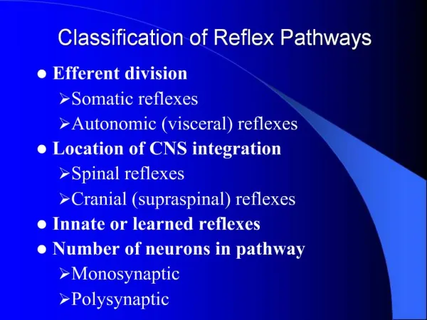
Reflexes • Reciprocal inhibition – cannot flex and extend the same muscle • Myotatic (knee-jerk) • Opposes gravity • Uses spindle sensory feedback • Reverse myotatic (knife-clasp) • Relaxes overloaded muscle • Responds to golgi tendon organ feedback
More Reflexes • Flexor reflex – response to pain • Crossed-extensor reflex – compensates for flexor reflex • One side extends as the other flexes • The circuit for coordinated control of walking resides in the spinal cord. • Circuits called “central pattern generators” give rise to rhythmic motor activity.