Download

1 / 42
420 likes | 623 Vues
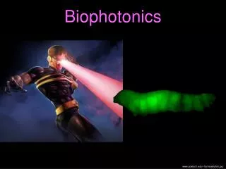
Biophotonics lecture 11. January 2012. Today: Correct sampling in microscopy Deconvolution techniques. Correct Sampling. Intensity [a.u.]. What is SAMPLING?. X [µm]. 1. 2. 3. 4. 5. 6. Intensity [a.u.]. 2. 3. 4. 5. 6.

E N D
Today: • Correct sampling in microscopy • Deconvolution techniques
Intensity [a.u.] What is SAMPLING? X [µm] 1 2 3 4 5 6
Intensity [a.u.] 2 3 4 5 6 There are many sine-waves, SAMPLED with the same measurements.Which is the correct one? Aliasing … suppose it is a sine-wave
Intensity [a.u.] 2 3 4 5 6 When sampling at the frequency of the signal, a zero-frequency is recorded! X [µm]
Intensity [a.u.] 2 3 4 5 6 X [µm]
Intensity [a.u.] 2 3 4 5 6 Problem:too high frequencies will be aliased, they will seemingly become lower frequencies X [µm]
Intensity Spatial Coordinate OTF Object: But … high frequencies are not transmitted well. Microscope Image: Intensity Spatial Coordinate
½ SamplingFrequency Aliased Frequencies Fourier-transform of Image Aliasing in Fourier-space SamplingFrequency Intensity NyquistRate Cut-off frequency=½ Nyquist Rate
Convolution of pixel form factor with sample Multiplication in Fourier-space Reduced sensitivity at high spatial frequency Intensity [a.u.] Pixel sensitivity X [µm] 1 2 3 4 5 6
rectangle form-factor OTF sampled contrast Optical Transfer Function 1 0 |kx,y| [1/m] Cut-off limit
Confocal: high Zoom morebleaching? Consequences of high sampling No! iflaserisdimmedor scan-speed adjusted badsignaltonoiseratio? Yes, but photonpositionsareonlymeasuredmoreaccuratelybinning still possiblehigh SNR. Readoutnoiseis a problemat high spatialsampling (CCD)
Multiplied in real spacewith band-limited information Reciprocal d-Sampling Grid Real-space sampling: Regular sampling
Reciprocald-Sampling Grid Real-space sampling: Regular sampling
In-Plane sampling distance • Axial sampling distance Widefield Sampling
In-Plane sampling distance (very small pinhole) • elseusewidefieldequation • Axial sampling distance Confocal Sampling
in-plane, in-focus OTF1.4 NA Objective Confocal OTFs WF Limit 1 AU 0.3 AU WF
Reciprocal d-Sampling Grid Multiplied in real spacewith band-limited information Real-space sampling: Hexagonal sampling Advantage: ~17%+ less ‚almostempty‘ informationcollected+ lessreadout-noiseapproximation in confocal
63× 1.4 NA Oil Objective (n=1.516), excitationat 488 nm, emissionat 520 nm leff = 251.75 nm, a = 67.44 deg widefield in-plane: dxy < 92.8 nm maximal CCD pixelsize: 63×92.8 = 5.85 µm confocal in-plane: dxy < 54.9 nm widefield axial: dz < 278.2 nm confocal axial: dz < 134.6 nm Fluorescence Sampling Example
OTF is not zero but very small(e.g. confocal in-plane frequency) Object possesses no higher frequencies You are only interested in certain frequencies (e.g. in counting cells, serious under-sampling is acceptable) Reasons for undersampling
If you need high resolution or need to detect small samples sample your image correctly along all dimensions Sampling Summary
Fluorescence imagingSample: S(r)Point SpreadFunction, PSF: h(r)Ideal Image: (Convolution operator ⨂)But: noisy image M(r) = N(M(r)) = E(r) + n(r) Poisson Noise
Naïve approach to deconvolution ?Problems:Fourier space: , Frequencies , for whichFourier space: Noise amplificationforlow
Poisson distributionProbability p formeasuring M photonswhenexpectationvalueis E photons: Image: http://en.wikipedia.org
Poisson probability in images Probability p formeasuringimage M withpixelvalues M(r) whenexpectationimage E withexpectationpixelvalues E(r): (Probabilities multiply) Or even:
Our goal: For a givenmeasurementimage M, find themostlikely sample distribution S. We can calculate: and But…
Bayes rule: But rather: The prior(requirespriorknowledge; canimplycontraints, e.g. positivity) Constant normalisationfactor
Nevertheless: Maximum likelihooddeconvolutiontriestomaximiseratherthan (uniform prior). The approach: Take thenegative naturallogartihmandminimise. Constant, therefore obsolete
Iterative minimisation: Simple “steepest gradient” search: Minimise function F(x) iteratively: with small Applied to log-likelihood function: With:
Richardson-Lucy: Steepestgradient Richardson Lucy (fix point iteration) Has positivity constraint!
Richardson-Lucy: Start withinitialguess: Problem withalgorithm: Veryslow Not stable
VirtualMicroscopy NO! FT Information & Photon noise Only Noise? 10 Photons / Pixel
Object Band Extrapolation? Mean Error Energy Relative Energy Regain Mean Energy
White Noise Object Is this always possible?
Is this always possible? Unfortunately NOT !