Download
1 / 23
230 likes | 406 Vues
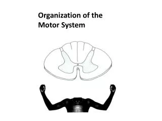
Organization of Motor Systems. The motor systems encompass 2 divisions of the PNS . Somatic (‘voluntary’) – controls skeletal muscle Autonomic (‘involuntary’) – controls visceral effectors: Smooth muscle surrounding blood vessels Cardiac muscle GI tract smooth muscle

E N D
The motor systems encompass 2 divisions of the PNS • Somatic (‘voluntary’) – controls skeletal muscle • Autonomic (‘involuntary’) – controls visceral effectors: • Smooth muscle surrounding blood vessels • Cardiac muscle • GI tract smooth muscle • Exocrine glands (salivary, sweat, gastric secretion, etc) • metabolic effects in variety of tissues (SNS)
Before we start, just a little about receptors • Receptors divided into two classes: • Ionotropic – receptor is an ion channel that opens a conductive pathway when bound with transmitter – effect may be depolarization, hyperpolarization or stabilization. • Metabotropic – transmitter-receptor binding initiates a chemical 2nd message within the target cell – this typically activates ion channels and transduces an electrical effect – but it doesn’t always have to do so to be effective. • Drugs that act on receptors may be • agonists (acting like or facilitating the action of the native transmitter) • antagonists (opposing the action of the native transmitter)
Are there excitatory and inhibitory transmitters and receptors? • Although we sometimes speak as if this were so, • In most cases, the outcome of applying a particular transmitter to a tissue depends entirely on what happens in the target tissue after the receptor has been activated, and not on any intrinsic property of the transmitter or receptor
Comparison of Autonomic and Somatic Motor Systems • The next slide compares the basic organization, transmitter identities, and receptor identities in the two branches of the autonomic system with the somatic system.
Some Important things to know about the motor systems slide • In all systems, at the 1st synapse outside the CNS, the transmitter is always Ach and the receptors nicotinic. • Nicotinic receptors are always excitatory; muscarinic and adrenergic receptors may be excitatory or inhibitory depending on the effect of the 2nd messengers coupled to the receptor in each tissue. • Single innervation of effectors in the somatic system vs (typically) dual innervation in the autonomic system. • Sympathetic ganglia are generally remote from the target organ whereas parasympathetic ones are generally in or on the target.
The nature of effectors in the autonomic system • Target tissues may include smooth muscle, ducted glands, and, in the case of the sympathetic branch, the metabolic activity of a number of tissues, including the liver, adipose tissue, skeletal muscle, and the brain itself. • Generally, autonomic inputs do not have a off/on effect on their targets. Instead, they modulate tissue activities that are already ongoing.
Smooth muscle as an effector of the autonomic system • Characteristics of smooth muscle: • Cells small • Surrounds hollow internal organs and blood vessels • Contractile machinery diffuse (no sarcomeres) • Electrical activation is dependent on external Ca++
Control features of smooth muscle • Two basic types: • 1. Single unit • Cells coupled by gap junctions • Spontaneous activity due to pacemakers • ANS does not need to make a synapse on each cell • ANS modulates spontaneous activity • 2. Multi-unit • Few gap junctions; little or no spontaneous activity • ANS must make synapses on each cell • Contractile activity reflects the balance of S versus PS inputs
There are two routes to activate smooth muscle Electromechanical: Chemical message > depolarization > Ca++ entry > contractile activity Pharmacomechanical: Chemical message > intracellular 2nd message > Ca++ release from internal stores > contractile activity
Anatomical summary of the ANS Basic Anatomical Layout of the Autonomic Nervous System
The Sympathetic Branch • Cell bodies of preganglionic neurons are in thoracic and upper lumbar cord • Preganglionic axons pass out through T1-L2 ventral roots as B and C fibers – small to very small. • They may synapse in paravertebral ganglion of the same segment, or turn to go rostrally or caudally to other segments, or continue through a splanchnic nerve to a prevertebral ganglion among the viscera • Celiac ganglion – organs of upper abdomen • Superior mesenteric ganglion – intestines • Inferior mesenteric ganglion – distal colon, bladder, genitalia
Sympathetic branch, continued. • Postganglionic axons are unmyelinated C fibers that may pass through gray ramus to spinal nerve running to body wall, or into a sympathetic nerve to abdominal organs • Adrenal medulla – preganglionics are in T10 and T11 – they go through the celiac ganglion to adrenal medulla where they synapse on chromaffin cells.
The Parasympathetic Branch • Preganglionic cell bodies are in brainstem nuclei • associated with particular cranial nerve roots, or • in S2-S4. • Ganglionic synapses are typically in or at least close to the target organs. • Cranial: ciliary ganglion – Cranial N. III; sphenopalatine and submaxillary ganglia – C. N. VII, Otic ganglion – C.N. IX • Abdomen - splenic flexure of colon marks the boundary between turf of the vagus N. (C.N. X) and the sacral part of the P.S.B.
The cholinergic synapse • Presynaptic: acetylcholine is synthesized from glucose • Postsynaptic: receptors may be nicotinic (ionotropic-excitatory) or muscarinic (2nd messenger); there are at least 4 subtypes of muscarinic receptors • After release, Ach is degraded by acetylcholinesterase and choline is reabsorbed by presynaptic terminal.
Cholinergic synapses as drug targets • Vesicle release blocker: botulinum toxin • Nicotinic receptor blockers: curare, cobra toxin, hexamethonium • Muscarinic receptor blockers: muscarine (toxin from fly agaric mushroom Amantia muscarina), atropine and scopalomine (Belladonna alkaloids) • Cholinesterase inhibitors: physostigmine, neostigmine, nerve gases, insecticides • Choline reuptake inhibitor: hemicholinium
The Adrenergic Synapse • Norepinephrine synthesized by synaptic terminal: • Tyrosine > DOPA>Dopamine>NE Norepinephrine is recovered by the presynaptic cell and may be recycled or degraded (monoamine oxidase)
Adrenergic synapse,cont. • 4 main classes of adrenergic receptors, characterized by agonist specificity • Alpha 1: sweat glands, blood vessels • Alpha 2: GI tract, presynaptic terminals • Beta 1: heart muscle • Beta 2: Lung airway; blood vessels in heart, skeletal muscle
The adrenergic synapse as a drug target • Alpha agonists: phenylephrine, pseudephedrine, amphetamines (decongestants, stimulants) • Alpha blockers: phentolamine, phenoxybenzamine, ergot alkaloids, yohimbine • Beta agonist: isoproterenol • Beta blockers: propranolol and its congeners (antihypertensives) • Monoamine oxidase inhibitors (antidepressants)
Higher Centers of Autonomic Function • Location: Brainstem, hypothalamus and other parts of the limbic system • Physiological variables controlled by the limbic system and A.N.S.: • Body temperature • Blood pressure • Blood glucose • Digestive activities • Sex response • Emotions
An example – baroreceptor reflex and blood pressure regulation Medullary Respiratory Center Limbic System Medullary CV center (brainstem) Vagus (inhibits) Sympathetics (stimulate) Baroreceptors (carotid sinus, aortic arch) Heart Vasculature
Reflexive response to a drop in blood pressure versus the medullary setpoint • Sensed by baroreceptors • Integrated in MCV center • Results of reflex activation: • Increases outflow of action potentials in thoracic sympathetics to heart • Heart rate rises • Stroke force increases • decreases or shuts off vagal outflow to heart • Parasympathetic tone removed – facilitates rate increase • Increases sympathetic outflow to blood vessels • Arterioles - Peripheral resistance rises • Veins - Venous capacitance falls, so blood moves to arterial side of circulation.