Download
1 / 12
120 likes | 228 Vues
This project focuses on creating a new set of detailed dosimetry models for pregnant females at three gestational stages (3, 6, and 9 months). Given the high radio-sensitivity of fetuses and the increasing need for precise dosimetry in nuclear medicine and radiation treatments involving pregnant patients, accurate models are essential. The work involves extracting 3D representations from CT images, adjusting organ masses to align with ICRP references, and studying the effects of various radiation sources, both internal and external. This effort aims to develop a standard set of phantoms for better safety in medical practices involving pregnant women.

E N D
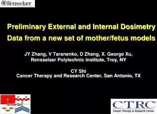
Preliminary External and Internal Dosimetry Data from a new set of mother/fetus models JY Zhang, V Taranenko, D Zhang, X. George Xu, Rensselaer Polytechnic Institute, Troy, NY CY Shi Cancer Therapy and Research Center, San Antonio, TX
Project Motivation(Pregnant Female Models) • High radio-sensitivity for the fetus • Needs for accurate dosimetry • Occupational (if the pregnancy is declared) • Nuclear medicine • Radiation treatment of pregnant patients (increasing!) • Air traveling • non-ionizing radiation (RF etc) • Models difficult to develop • Twice as many tissues and organs • 3-month, 6-month and 9-month gestational periods • Medical images are rare
Project Goals 1) Development a new set of models of pregnant mother and fetus at the end of 3-, 6- and 9-month gestational periods 2) Compile organ dose parameters for external and internal irradiations
Existing Pregnant Female Models • Stylized models • Stabin 1995 • Chen 2004 From partial-body CT image set - Shi and Xu (2004) They are un-realistic and in-complete
Method:Flow Chart of Pregnant Female Model Development Model Extraction of 3D representation from CT Images (external uterine wall). This is a new approach!
From Surfaces to Voxels Re-voxelize at any size (1mm shown here) Put into MC codes - MCNPX - EGSnrc Surface Model Adjusted to ICRP reference values Geometry in MCNPX code Voxel Model
RPI-P3, RPI-P6, and RPI-P9 Models 9-month 3-month 6-month
Results - Internal photon Specific Absorbed Fractions(RPI-P9 phantom)
Conclusion • A series of pregnant female and fetus phantoms have been developed • Organ masses are adjusted according to ICRP reference values • External photon, electron, neutron and proton sources have been studied • Internal photon and electron sources have been studied • ICRP is hoped to adopt the RPI-P series as standard pregnant-female models
Acknowledgements • This work is supported by grants 1R42CA115122-01 and 5R01CA116743-03 from the National Institutes of Health • Drs Mike Stabin and Keith Eckerman provided valuable help on • ICRP data analysis Visualization for RPI-P9 Pregnant Female Model (available at RRMDG.rpi.edu)