Download
1 / 17
180 likes | 366 Vues
Influence of Backscattering on the Spatial Resolution of Semiconductor X-ray Detectors. Martin Hoheisel Siemens Medical Solutions, Forchheim, Germany Alexander Korn, Jürgen Giersch Institute of Physics, University Erlangen/Nürnberg, Germany

E N D
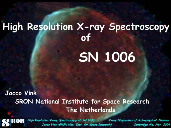
Influence of Backscatteringon the Spatial Resolutionof Semiconductor X-ray Detectors Martin Hoheisel Siemens Medical Solutions, Forchheim, Germany Alexander Korn, Jürgen Giersch Institute of Physics, University Erlangen/Nürnberg, Germany 6th International Workshop on Radiation Imaging Detectors Glasgow, Scotland, UK, 2004
Outline • Introduction • What limits spatial resolution? • Simulations with and without backscattering objects • Results • Summary and conclusions
Introduction • Medical X-ray detectors • Past: film/screen combination, image intensifier, storage phosphor • Future: semiconductor-based flat-panel detector • Registration concepts • Scintillator + matrix of photodiodes and switches • Directly absorbing semiconductor + matrix of switches • Signal integration or counting of X-ray quanta • Spatial resolution required • Soft tissue: 0 … 2 lp/mm (line pairs per mm) • Bones: 0 … > 3 lp/mm • Dental, mammography: 0 … > 5 lp/mm … 10 lp/mm … 20 lp/mm
What limits spatial resolution? • X-ray interaction with absorber • Elastic (Rayleigh) scattering • Inelastic (Compton) scattering generating fast electrons • Photoelectric absorption generating fluorescent quanta and fast electrons • Electron energy loss • Elastic scattering • Multiple excitation of electron-hole pairs Primary X-ray quanta Compton scatter Photoelectric effect X-ray Fluorescence X-ray absorber Electron energy loss Rayleigh scatter Backscattered quanta X-ray absorption, energy loss, i.e. signal generation are distributed in space Scattering object
e- What else limits spatial resolution? • Transport (for directly absorbing detectors) • Charge carriers are collected by drift • Charge carriers are spread by diffusion • Integration and sampling • Signal is integrated over pixel area • Ambiguity by sampling for spatial frequencies > Nyquist frequency Simple model: Transit time Diffusion constant Diffusion length independent of material properties! + Diffusion Drift – Lateral spread
Simulations • Monte Carlo simulation • Program, ROSI (Roentgen Simulation), based on EGS4 algorithm • Mono-energetic range 20 keV … 120 keV • Common spectra 28 kV … 120 kV • X-ray fan beam hits a pixelated detector grid • Slightly (5°) tilted to produce oversampling • Line spread function • Used to calculate the modulation transfer function (MTF)
10 µm Setup (1) • Materials and detectors investigated • 300 µm and 700 µm Si on Medipix-2 (55 µm pixels) • 300 µm GaAs on Medipix-2 (55 µm pixels) • Setup • Absorbing semiconductor layer300/700 µm Si (300 µm GaAs) • Bump-bond level1.8 µm In layer(bumps 21.8 µm in diameter) • Medipix-2 chip500 µm Si • Printed circuit board1 mm PMMA 20 (60) µm
1 µm 1 µm Setup (2) • Materials and detectors investigated • 200 µm Se on a-Si readout matrix (70 µm pixels) • 420 µm CsI on a-Si readout matrix (143 µm pixels) • Setup • Absorbing layer200 µm Se (420 µm CsI) • Glass substrate3 mm Corning 7059 • Protective shield1 mm Pb
Results (1) • 20 keV ... 120 keV • 300 µm Si with Medipix-2 and chip board • Low-frequency drop visible for E > EK(In) = 27.94 keV
Results (2) • 300 / 700 µm Si with Medipix-2 and chip board at 50 keV • Si without backscattering objects behind the absorber • In bumps or homogeneous layer • Low-frequency drop of MTF is due to backscatter
Results (3) • 300 / 700 µm Si with Medipix-2 and chip board at 120 keV • Backscattered quanta from chip and chipboard are partly absorb-ed by the In layer, but penetrate the gaps between the bumps • The thinner Si leads to a stronger backscatter effect
Results (4) • 200 µm Se layer with a-Si readout matrix on glass substrate (3 mm Corning 7059) and additional 1 mm Pb shielding • Low-frequency drop caused by backscatter (compare to inset) without backscatter
Results (5) • 420 µm CsI layer with a-Si readout matrix on glass substrate (3 mm Corning 7059) and additional 1 mm Pb shielding (optical effects on MTF not taken into account) • Low backscattering has low effect on MTF (compare to inset) without backscatter
Scatter fraction • Scatter fraction rises above K-edge of backscattering object • Indium bumps for Medipix-2, i.e. E > EK(In) = 27.94 keV • Glass substrate for CsI and Se detectors, i.e. E > EK(Ba) = 37.44 keV
Summary • Upper limit for MTF is the sinc function of the pixel size • At energies below K-edge, the MTF is close to the sinc function • At energies above K-edge, fluorescence spreads the signal, thus reducing resolution • Above 50 keV, resolution is reduced by the range of fast electrons • Carrier transport has an influence on MTF • Transmitted and backscattered quanta reach the whole detector • MTF is reduced at low spatial frequency low-frequency drop • Effect increases above K-edge of backscattering object • Monochromatic quanta and common X-ray spectra lead to comparable results
Conclusions • The effect of backscatter on spatial resolution must not be ignored • Important with thin absorbers, where a high fraction of incident quanta is transmitted • Dependent on materials behind absorber (substrate, chips) • Interpretation of charge sharing may be wrong due to backscattered quanta • Can be tested by experiments below/above K-edge