Download
1 / 26
450 likes | 2.14k Vues
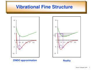
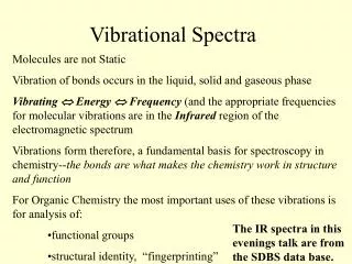
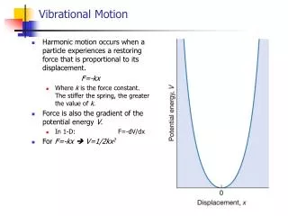
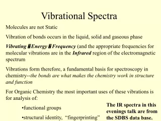
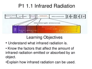
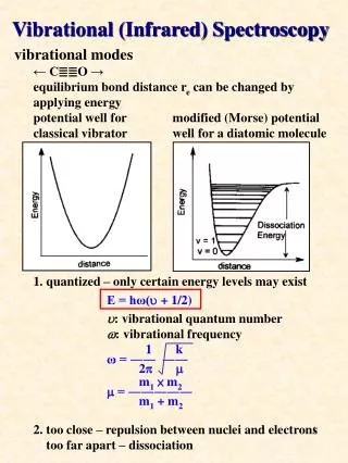
Vibrational (Infrared) Spectroscopy. vibrational modes ← C≣≣O → equilibrium bond distance r e can be changed by applying energy potential well for modified (Morse) potential classical vibrator well for a diatomic molecule

E N D
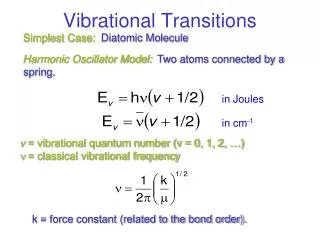
Vibrational (Infrared) Spectroscopy vibrational modes ← C≣≣O → equilibrium bond distance re can be changed by applying energy potential well for modified (Morse) potential classical vibrator well for a diatomic molecule 1. quantized – only certain energy levels may exist E = hω(u + 1/2) u: vibrational quantum number w: vibrational frequency 1 k ω = ―― ―― 2p m m1× m2 m = ――――― m1 + m2 2. too close – repulsion between nuclei and electrons too far apart – dissociation
ex. HCl u(HCl) = 2990 cm-1 DCl u(DCl) = 2145 cm-1 ex. u(NO) bond order NO+ 2273 cm-1 3 NO 1880 cm-1 2.5 NO- 1365 cm-1 2 NO2- 886 cm-1 1.5 number of vibrational modes a molecule consists of N atoms, there are 3N degrees of freedom translation rotation vibration nonlinear 3 3 3N – 6 linear 3 2 3N – 5 type of vibrational modes stretching modeubending moded IR active absorption Raman active absorption
frequencies for some commonly encountered groups, fragments, and linkages in inorganic and organic molecules
ex. W(CO)6 Mn(CO)5Br compound u(CO) (cm-1) [Ti(CO)6]2- 1740 [V(CO)6]- 1860 Cr(CO)6 2000 [Mn(CO)6]+ 2095 stretching modes of CO and IR frequencies (a) terminal (b) doubly bridging (c) triply bridging ex.
some ligands capable of forming linkage isomers IR spectrum for nujol salt plates NaCl 625 cm-1 KBr 400 cm-1 CsI 200 cm-1 721 1377 1462 2925 2855
symmetry of normal vibrations ex. CO32- 6 vibrational modes C3(u3a) = -1/2u3a + 1/2 u3b C3(u3b) = -3/2u3a - 1/2 u3b determine the symmetry type of normal modes E c = 12
C3 c= 0 C2 c = -2 c (sh) = 4 c (S3) = -2 ?? c (sv) = 2
G = A1’ + A2’ + 3E’ + 2A2” + E” 3 translatory modes: E’, A2” 3 rotational modes: A2’, E” genuine vibrational modes: Gg = A1’ +2E’ + A2” IR active: E’, A2” (3 bands) Raman active: A1’, E’ (3 bands) particular internal coordinates to normal modes C—O bonds E C3 C2sh S3sv G 3 0 1 3 0 1 GCO = A1’ + E’ in-plane stretching GOCO = A1’ + E’ in-plane bending A2” out-of-plane bending ex. determine the number of IR active CO stretching bands for the following metal carbonyl compounds : M(CO)6 M(CO)5L cis-M(CO)4L2 trans-M(CO)4L2fac-M(CO)3L3 mer-M(CO)3L3 M(CO)5 M(CO)4L M(CO)3L2 M(CO)4
(i) trans-M(CO)4L2 D4h E C4 C2 C2’ C2” i S4shsvsd L 4 0 0 2 0 0 0 4 2 0 OCCO OC CO==> A1g + B1g + Eu L IR-active: Eu (ii) cis-M(CO)4L2 COC2v E C2ss’ OCL4 0 2 2 OC L==> 2A1 + B1 + B2 CO IR-active: 2A1, B1, B2 (iii) mer-M(CO)3L3 COC2v E C2ss’ L L 3 1 1 2 OC L ==> 2A1 + B1 OC IR-active: 2A1, B1 (iv) M(CO)5 D3h E C3 C2sh S3sv 5 2 1 3 0 3 ==> 2A1’ + A2” + E’ IR-active: A2”, E’
(v) M(CO)4L LD3v E C3sv 4 1 2 ==> 2A1 + E IR-active: 2A1, E D2v E C3svsv‘ L 4 0 2 2 ==> 2A1 + B1 + B2 IR-active: 2A1, B1, B2 (vi) M(CO)3L2 LD3h E C3 C2sh S3sv 3 0 1 3 0 1 ==> A1‘ + E’ L IR-active: E’ LCs E sh 3 1 L ==> 2A‘ + A” IR-active: 2A’, A” (vii) M(CO)4 Td E C3 C2 S3sd 4 1 0 0 2 ==> A1 + T2 IR-active: T2
number of CO stretching bands in IR spetcrum for metal carbonyl compounds
calculation of force constants for diatomic molecule AB harmonic oscillator f • m-1 – l = 0 for polyatomic molecule Wilson’s method “The F and G matrix method” |FG – El| = 0 F: matrix of force constant (potential energy) G: matrix of masses and spatial relationship of atoms (kinetic energy) E: unit matrix e.g. H2O Gg = 2A1 + B1 2 O-H distance Dd1, Dd2 A1 + B1 ∠HOH DθA1 using projection operator to obtain complete set of symmetry coordinates for vibrations A1 : S1 = Dθ S2 = 1/√2(Dd1 + Dd2) B1 : S3 = 1/√2(Dd1 - Dd2) F matrix 2V = Sfik si sk si, sk: change in internal coordinates for Dd1Dd2Dθ Dd1 fd fdd fdθ Dd2 fdd fd fdθ Dθ fdθ fdθ fθ
2V = fd(Dd1)2 + fd(Dd2)2 + fθ(Dθ)2 + 2 fdd(Dd1Dd2) + 2 fdθ(Dd1Dθ) + 2 fdθ(Dd2Dθ) =[Dd1Dd2Dθ] fd fdd fdθ Dd1 fdd fd fdθ Dd2 fdθfdθfθDθ = s’f s relationship between the internal coordinates and the symmetry coordinates S = U s U matrix Dq 0 0 1 Dd1 Dd1 + Dd2 = 1/√2 1/√2 0 Dd2 Dd1 - Dd2 1/√2 -1/√2 0 Dq S = U s s = U’ S s’ = (U’ S)’ = S’U s’fs = S’FS (S’U)f(U’S) = S’FS S’(UfU’)S = S’FS ==> F = UfU’ 0 0 1 fd fdd fdθ 0 1/√2 1/√2 F =1/√2 1/√2 0 fdd fd fdθ 0 1/√2 -1/√2 1/√2 -1/√2 0 fdθ fdθ fθ 1 0 0 fθ √2 fdθ 0 = √2 fdθfd+ fdd 0 0 0 fd - fdd
G matrix G = UgU’ 0 0 1 gd gdd gdθ 0 1/√2 1/√2 G = 1/√2 1/√2 0 gdd gd gdθ 0 1/√2 -1/√2 1/√2 -1/√2 0 gdθ gdθ gθ 1 0 0 g33 √2 g13 0 = √2 g13 g11 + g12 0 0 0 g11 - g12 g11 = mH + mO g12 = mO cosθ g13 = -(mO/r) sinθ g33 = 2(mH + mO - mO cosθ)/r2 m : reciprocal of the mass 2(mH + mO - mO cosθ)/r2 -(√2mO/r) sinθ 0 G = -(√2mO/r) sinθ mH + mO (1+ cosθ) 0 0 0 mH + mO (1 - cosθ) for H2O θ= 104.3o31’ r = 0.9580 Å 2.332 -0.0893 0 G = -0.0893 1.0390 0 0 0 1.0702 fθ √2 fdθ 2.332 -0.0893 l 0 A1: – = 0 √2 fdθ fd+ fdd -0.0893 1.0390 0 l B1: 1.0702(fd - fdd)= l
elements of the g matrix mi: reciprocal mass of the ith atom rij: reciprocal of the distance between ith and jth
Raman spectroscopy light of energy less than that required to promote a molecule into an excited electronic state is absorbed by a molecule, a virtual excited state is created virtual state is very short lifetime, the majority of the light is re-emitted over 360oC, this is called Rayleigh scattering C. V. Raman found that the energy of a small proportion of re-emitted light differs from the incident radiation by energy gaps that correspond tosome of the vibrational modes Stokes lines anti-Stokes line
schematic representation of Raman spectrometer selection rules for vibrational transitions •a fundamental will be infrared active if the normal mode which is excited belongs to the same representation as any one or several of the Cartesian coordinates •a fundamental will be Raman active if the normal mode involved belongs to the same representation as one or more of the components of the polarizability tensor of the molecule the exclusion rule – in centrosymmetric molecules, no Raman-active vibration is also IR-active and no IR-active vibration is also Raman-active only fundamentals of modes belonging to g representations can be Raman active and only fundamentals of modes belonging to u representations can be IR active
ex. Na2MoO4 dissolved in HCl exhibits Raman peaks at 964, 925, 392, 311, 246, 219 cm-1 925, 311 cm-1 being polarized what can be deduced from the spectrum? no n(Mo—H) and n(O—H) bands only M—Cl and M=O likely exist 964, 925 cm-1 Mo=O stretching bands 392 cm-1 Mo=O bending mode 311, 246, 219 cm-1 Mo—Cl stretching modes possible product: