Download
1 / 59
770 likes | 1.52k Vues
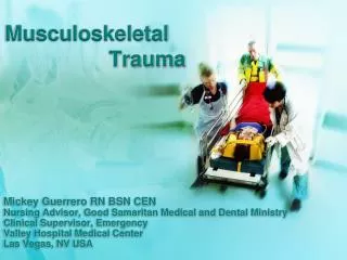
Chapter 69 Management of Patients With Musculoskeletal Trauma. Injuries of the Musculoskeletal System. Contusion: soft tissue injury produced by blunt force Pain, swelling, and discoloration: ecchymosis Strain: pulled muscle-injury to the musculocutaneous unit

E N D
Chapter 69Management of Patients With Musculoskeletal Trauma
Injuries of the Musculoskeletal System • Contusion:soft tissue injury produced by blunt force • Pain, swelling, and discoloration: ecchymosis • Strain: pulled muscle-injury to the musculocutaneous unit • Pain, edema, muscle spasm, ecchymosis, and loss of function are on a continuum graded 1st , 2nd, and 3rd degree
Injuries of the Musculoskeletal System (cont.) • Sprain: injury to ligaments and supporting muscle fiber around a joint • Joint is tender and movement is painful; edema, disability, and pain increase during the first 2 to 3 hours • Dislocation: articular surfaces of the joint are not in contact • A traumatic dislocation is an emergency with pain change in contour, axis, and length of the limb and loss of mobility
Common Sports-Related Injuries • Contusions, strains, sprains, and dislocations • Tendonitis: inflammation of a tendon by overuse • Meniscal injuries of the knee occur with excessive rotational stress • Traumatic fractures • Stress fractures
Prevention of Sports-Related Injuries • Use of proper equipment: running shoes for runners, wrist guards for skaters, etc. • Effective training and conditioning specific for the person and the sport • Stretching prior to engaging in a sport or exercise has been recommended but may not prevent injury • Changes in activity and stresses should occur gradually • Time to “cool down” • Tune in to the body; be aware of limits and capabilities • Modify activities to minimize injury and promote healing
Occupational-Related Injuries • Common injuries include strains, sprains, contusions, fractures, back injuries, tendonitis, and amputations • Prevention measures include personnel training, proper use of equipment, availability of safety and other types of equipment (patient lifting equipment, back belts), correct use of body mechanics, and institutional policies
Types of Fractures • Complete • Incomplete • Closed or simple • Open or compound/complex • Grade I • Grade II • Grade III
Manifestations of Fracture • Pain • Loss of function • Deformity • Shortening of the extremity • Crepitus • Local swelling and discoloration • Diagnosis by symptoms and x-ray • Patient usually reports an injury to the area
Emergency Management • Immobilize the body part • Splinting: joints distal and proximal to the suspected fracture site must be supported and immobilized • Assess neurovascular status before and after splinting • Open fracture: cover with sterile dressing to prevent contamination • Do not attempt to reduce the fracture
Medical Management • Reduction • Closed • Open • Immobilization: internal or external fixation • Open fractures require treatment to prevent infection • Tetanus prophylaxis, antibiotics, and cleaning and debridement of wound • Closure of the primary wound may be delayed to permit edema, wound drainage, further assessment, and debridement if needed
Nursing Management of the Patient With a Simple Fracture • Assessment: include neurovascular assessment, pain, activity limitations, patient knowledge, and home environment and support • Goal is to have patient return to usual activities as soon as possible • Patient teaching is a primary intervention as the patient will usually be cared for in the home setting • See Chart 69-2
Complications of Fractures • Factors that affect fracture healing: see Chart 69-3 • Shock • Fat embolism • Compartment syndrome • Delayed union and nonunion • Avascular necrosis • Reaction to internal fixation devices • Complex regional pain syndrome (CRPS) • Heterotrophic ossification
Rehabilitation Related to Specific Fractures • Clavicle • Use of claviclar strap (“figure 8”) or sling • Exercises • Limitation of activities • Do not elevate arm above shoulder for approximately 6 weeks • Humeral neck and shaft fractures • Slings and bracing • Activity limitations and pendulum exercises
Rehabilitation Related to Specific Fractures • Elbow fractures • Monitor regularly for neurovascular compromise and signs of compartment syndrome • Consider potential for Volkmann's contracture: see Chart 69-4 • Encourage active exercises and ROM to prevent limitation of joint movement after immobilization and healing (4 to 6 weeks for nondisplaced, casted) or after internal fixation (about 1 week)
Rehabilitation Related to Specific Fractures (cont.) • Colles’ fracture • Early functional rehabilitation exercises • Active motion exercises of fingers and shoulder • Pelvic fractures • Management depends upon type and extent of fracture and associated injuries • Stable fractures are treated with a few days’ bed rest and symptom management • Early mobilization reduces problems related to immobility
Rehabilitation Related to Specific Fractures (cont.) • Hip fracture • Surgery is usually done to reduce and fixate the fracture • Care is similar to that of a patient undergoing other orthopedic surgery or hip replacement surgery
Rehabilitation Related to Specific Fractures • Femoral shaft fractures • Lower leg, foot, and hip exercises to preserve muscle function and improve circulation • Early ambulation stimulates healing • Physical therapy, ambulation, and weight bearing are prescribed • Active and passive knee exercises are begun as soon as possible to prevent restriction of knee movement
Rehabilitation Related to Specific Fractures (cont.) • Uncomplicated rib fractures • Chest strapping is not used • Encouraged to cough and deep breathe
Rehabilitation Related to Specific Fractures • Thoracolumbar spine fractures • Usually treated conservatively with limited bed rest • Avoid sitting • Progressive ambulation • Emphasize good posture and body mechanics • Implement back strengthening exercises
Nursing Process—Assessment of the Patient With Fracture of the Hip • Health history and presence of concomitant problems • Pain • VS, respiratory status, LOC, and signs and symptoms of shock • Affected extremity including frequent neurovascular assessment • Bowel and bladder elimination, bowel sounds, and I&O • Skin condition • Anxiety and coping
Nursing Process—Diagnosis of the Patient With Fracture of the Hip • Acute pain • Impaired physical mobility • Impaired skin integrity • Risk for impaired urinary elimination • Risk for ineffective coping • Risk for disturbed thought processes
Collaborative Problems/Potential Complications • Hemorrhage • Peripheral neurovascular dysfunction • DVT • Pulmonary complications • Pressure ulcers
Nursing Process—Planning the Care of the Patient With Fracture of the Hip • Major goals include pain relief; achievement of a pain-free, functional, and stable hip; healed wound; maintenance of normal urinary elimination pattern; use of effective coping mechanisms; an oriented patient who participates in decision making; and absence of complications
Relief of Pain • Administer analgesics as prescribed • Use of Buck’s traction as prescribed • Handle extremity gently • Support extremity with pillows and when moving • Position for comfort • Provide frequent position changes • Provide alternative pain relief methods
Prompting Physical Mobility • Maintain neutral position of hip • Use trochanter rolls • Maintain abduction of hip • Implement isometric, quad-setting, and gluteal- setting exercises • Use trapeze • Use ambulatory aids • Consult with physical therapy
Interventions • Use aseptic technique with dressing changes • Avoid/minimize use of indwelling catheters • Support coping • Provide and reinforce information • Encourage the patient to express concerns • Support coping mechanisms • Encourage the patient to participate in decision making and planning • Consult social services or other supportive services
Interventions (cont.) • Orient patient to and stabilize the environment • Provide for patient safety • Encourage participation in self-care • Encourage coughing and deep breathing exercises • Ensure adequate hydration • Apply TED hose or SCDs as prescribed • Encourage ankle exercises • Provide patient and family teaching
Rehabilitation of Patients With Amputation • Amputation may be congenital, traumatic, or due to conditions such as progressive peripheral vascular disease, infection, or malignant tumor • Amputation is used to relieve symptoms, improve function, and save the person's life • The health care team needs to communicate a positive attitude to facilitate acceptance and participation in rehabilitation
Rehabilitation Needs • Psychological support • Prosthesis fitting and use • Physical therapy • Vocational/occupational training and counseling • Use a multidisciplinary team approach • Patient teaching: seeChart 69-6