Download
1 / 42
480 likes | 967 Vues
Image Gently, Pause and Pulse: Practice of ALARA in Pediatric Fluoroscopy. Sue C. Kaste, DO 1, 2 Marta Hernanz-Schulman, MD 3 Ishtiaq H. Bercha, M.Sc. 4 1 St. Jude Children’s Research Hospital 2 University of Tennessee Health Science Center

E N D
Image Gently, Pause and Pulse: Practice of ALARA inPediatric Fluoroscopy Sue C. Kaste, DO1, 2 Marta Hernanz-Schulman, MD3 Ishtiaq H. Bercha, M.Sc. 4 1 St. Jude Children’s Research Hospital 2 University of Tennessee Health Science Center 3 Monroe Carell Jr. Children’s Hospital at Vanderbilt 4 The Children’s Hospital, Aurora, Colorado.
ALARA • “As Low As Reasonably Achievable” • General principle guiding radiation exposure • Keep exposure to radiation dose as low as is possible for each procedure, while obtaining needed clinical information • = Image Optimization
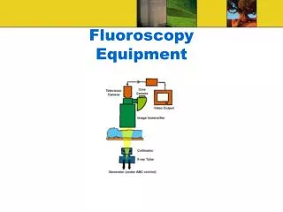
Primary Learning Objective • Review pediatric fluoroscopic procedures • understand the source of radiation • understand methods to reduce radiation • effect on image quality
Other Learning Objectives • Fluoroscopy radiation units. • Scope of pediatric fluoroscopic procedures • Methods available for dose reduction • clinical settings to apply dose reduction
Fluoroscopy Radiation Units Basic Radiation Quantities : • Exposure & Exposure Rate • Air Kerma & Air Kerma Rate
Fluoroscopy Radiation Units Radiation Measurement Quantities: • Incident Air Kerma & Rate • Entrance Surface Air Kerma & Rate
Fluoroscopy Radiation Units • Absorbed dose • Equivalent Dose • Effective dose • Risk Related Quantities:
Basic Radiation Quantities • Exposure – expresses intensity of x-ray energy per unit mass of air. Units: Coulomb per kilogram (C/kg). Commonly used units are Roentgen or milli Roentgen, expressed as R or mR, respectively. 1 R = 2.58 x 10⁻⁴ C/kg • Exposure rate identifies x-ray intensity per unit time. Commonly used units are R/min or mR/min.
Basic Radiation Quantities • Air Kerma (K) – sum of initial kinetic energies of all charged particles generated by uncharged particles such as x-ray photons released per unit mass of air. Unit = Joule per kilogram, Commonly referred to as Gray/milli Gray (Gy or mGy). 1 Roentgen of exposure 8.7 mGy air kerma • Air Kerma Rate quantifies air kerma per unit time and is written as, dK/dt, that is, incremental kerma per unit increment of time.
Measurement Quantities • Incident Air Kerma (Ka,i)– is the air kerma from the incident beam along the central x-ray beam axis at the skin entrance plane. • Only the primary beam is considered and the effect of back scattered radiation is excluded. Unit = Joule per kilogram, Commonly referred to as Gray/milli Gray (Gy or mGy). Incident Air Kerma Rate quantifies air kerma per unit time. It is usually measured as mGy/min.
Measurement Quantities • Entrance Surface Air Kerma (Ka,e) – It is the air kerma from the incident beam along the central x-ray beam axis at the point where radiation enters the patient and the effect of back scattered radiation is included. Given as Ka,e = Ka,i x B B = Back Scatter Factor. Unit = Joule per kilogram, Commonly referred to as Gray & milli Gray (Gy or mGy). Incident Air Kerma Rate quantifies air kerma per unit time.
Risk Related Quantities • Absorbed dose – energy deposited per unit mass of a material, in our case, within tissue. • Initially measured as rads • Current unit based on Systeme Internationale (SI unit) SI Unit of Absorbed Dose = Gray • 1Gray (Gy) = 100 rad • 1rad = 10 mGy
Risk Related Quantities • Dose Equivalent – accounts for biological effect of type of radiation • For example, difference in biological effect between • , and radiation • Radiation Weighting factor (wR) – scaling factor used • , Xray wR = 1 • (wR) = 20 • SI Unit is Sievert • 1 Sievert (Sv) = 100 rem • 1 rem = 10 mSv
Risk Related Quantities • Effective dose – accounts for radio-sensitivity of specific organs • Includes • A tissue weighting factor (wT) for each sensitive organ • Each tissue included in the clinical examination (HT) • Effective dose = wT x HT, () summed over all exposed organs. • SI Unit is Sievert • 1 Sievert (Sv) = 100 rem • 1 rem = 10 mSv
Background Radiation Exposure * = estimate at sea level in US
Medical Radiation Exposures * Ward et al Radiology 2008;249:1002
Practical Methods to Reduce Radiation Dose toFluoroscopy Staff &Patients
Reduce Radiation Dose: Staff • Staff dose is due to scattered radiation • Scattered radiation is directly proportional to Patient Dose Patient Dose Staff Dose
Staff Protection • Well fitted lead apron (knees) • Leaded glasses (with sides) • Thyroid shield • Lead gloves
Staff protection: Hands • Keep hands out of the beam • Collimate
Staff protection: Shields • Lead shield on tower • Do not turn your back to Xray beam if wearing front apron only
In summary: Have we…. • … left our hands in the beam? • … sacrificed personal safety for expediency? • … turned our unshielded backs to the X-ray source? • … unnecessarily prolonged exposure? • … pushed away a protective barrier?
Patient Protection • Radiation dose is optimized when we use • Least amount of radiation • That delivers clinically adequate image quality
Patient Positioning • Proper patient positioning • Make use of Inverse square law! • Maximize distance between x-ray tube & patient • Minimize distance between patient & Image Intensifier
Control Fluoroscopic Exposures • Choose pulsed fluoroscopy • Choose as short a pulse width as possible • Typically 5 – 10 msec pulse width
Control Fluoroscopic Exposures • Continuous fluoroscopy • 30 pulses per second • 33 msec pulse width • Grid-controlled fluoroscopy • e.g. 15 pulses / sec • 10 msec pulse width
Control Fluoroscopic Exposures • Increase filtration to reduce patient radiation dose • Balanced by need for shorter pulse widths to freeze motion • Interposition of Aluminum and variable thickness of Copper • Removes low energy radiation that does not reach the image intensifier • scattered within the patient • adds radiation dose • does not contribute to image
Control Fluoroscopic Exposures • Remove anti-scatter grid whenever possible • Removes scattered radiation • Increased radiation dose • Not necessary in small patients • Avoid unnecessary magnification
Control Fluoroscopic Exposures • Collimate to area of interest • No need to radiate tissue that is not clinically pertinent
Control Fluoroscopic Exposures • Use “last image hold” • Whenever you need to inspect the anatomy, and do not need to observe motion or changes with time • Use Fluoroscopy Store (FS) • this method is ideal to convey and record motion, such as peristalsis, or show viscus distensibility, as in esophagram • when you need information without excessive detail Exposure Fluoro-grab
Control Number of Images • Choose appropriate, patient-specific technique • Limit acquisition to what is essential for diagnosis and documentation • PAUSE– Plan study ahead • PAUSE- think # frames / second • PAUSE – think magnification • PAUSE – think Last Image Hold • PAUSE – think Image Grab
Control Fluoroscopic Use Use fluoroscopic examination when there is a clear medical benefit. Use alternative imaging methods whenever possible US MRI
Special Pediatric Considerations • Pediatric patient management more critical • Increased radio-sensitivity, small size, longevity. • Pediatric size • Smaller patient leads to less scattered radiation • There is an increased need for magnification
Institutional Strategies to Optimize Radiation Exposure Fluoroscopy
To Start: • An in-house diagnostic medical physicist in pediatric hospitals is optimal. • The physicist must have proper training and background in Medical Physics, such as CAMPEP accredited graduate and residency programs. • Proper training is key
To Start: An Image Management committee, comprised of radiologists, technologists, administrators and medical physicists, under the direction of the department Chair, can be very helpful. • Responsible for optimizing radiation procedures. • Oversee the departmental QA/QC program. • Meet criteria for accreditation, e.g. ACR
To Start: • Oversee purchase of capital equipment and periodic hardware and software upgrades. • Staff training on state of the art technologies. • Technologists, radiologists • Equipment, safety, physics, radiation biology • Compliance with applicable state and federal regulations.
Dosimetry Records • Manage fluoroscopy parameters • e.g., pulsed fluoroscopy, pulse rate, removable grid • Record information related to patient radiation dose as displayed by the equipment: • Cumulative Dose Area Product. • Cumulative Air kerma/Skin Dose.
Summary • PAUSEto properly plan and prepare for study • Activate dose saving features of equipment • No image exposures unless necessary • Download image grab instead • PULSEat lowest possible rate
References -Gelfand, D.W., D.J. Ott, and Y.M. Chen, Decreasing numbers of gastrointestinal studies: report of data from 69 radiologic practices. AJR Am J Roentgenol, 1987. 148(6): p. 1133-6. -Margulis, A.R., The present status and the future of gastrointestinal radiology. Abdom Imaging, 1994. 19(4): p. 291-2. -Page, M. and H. Jeffery, The role of gastro-oesophageal reflux in the aetiology of SIDS. Early Hum Dev, 2000. 59(2): p. 127-49. -Strauss KJ, Kaste SC. The ALARA (as low as reasonably achievable) concept in pediatric interventional and fluoroscopic imaging: striving to keep radiation doses as low as possible during fluoroscopy of pediatric patients—a white paper executive summary. Radiology 2006 240(3):621-622. -Ward VL, Strauss KJ, Barnewolt CE, Zurakowski D, Venkatakrishnan V, Fahey FH, Lebowitz RL, Taylor GA. Pediatric radiation exposure reduction and effective dose reduction during voiding cystourethrography. Radiology 2008 249:1002-1009. -Hall, E. and J. Amato, Radiobiology for the Radiologist. 2005: Williams & Wilkins. -Lederman, H.M., et al., Dose reduction fluoroscopy in pediatrics. Pediatr Radiol, 2002. 32(12): p. 844-8. -Ward, V., et al., Radiation exposure reduction during voiding cystourethrography in a pediatric porcine model of vesicoureteral reflux. Radiology, 2005. 235. -Boland, G.W.L., et al., Dose Reduction in Gastrointestinal and Genitourinary Fluoroscopy: Use of Grid-Controlled Pulsed Fluoroscopy. Am. J. Roentgenol., 2000. 175(5): p. 1453-1457. -Brown, P.H., et al., A multihospital survey of radiation exposure and image quality in pediatric fluoroscopy. Pediatr Radiol, 2000. 30(4): p. 236-42. -Strauss KJ. Pediatric interventional radiography equipment: safety considerations. Pediatr Radiol (2006) 36 (Suppl 2):126-135. -Hernanz-Schulman M, Emmons M, Price R. Radiation dose reduction and image quality considerations in pediatric patients. Radiology RSNA syllabus, November, 2006