Download
1 / 63
630 likes | 775 Vues
This paper explores the intricate structure and function of inositol trisphosphate receptors (IP3Rs) in regulating intracellular calcium dynamics. It highlights how IP3Rs, composed of multiple subunits, interact with various proteins, including regulatory proteins, kinases, and anchoring proteins, which modulate their activity and localization. The involvement of the N-terminal region in suppressing IP3 binding is discussed, along with the effects of calmodulin (CaM) on IP3R functionality. The findings provide insights into the physiological implications of IP3R modulation in cellular signaling processes.

E N D
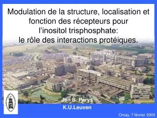
Modulation de la structure, localisation et fonction des récepteurs pourl’inositol trisphosphate: le rôle des interactions protéiques. Jan B. Parys K.U.Leuven Orsay, 7 février 2003
Intracellular Ca2+ homeostasis (Clapham, 1995)
Structure of the IP3R – the simplified view Ca2+ Cytoplasm N C Lumen of ER Lumen of ER • 4 subunits = 1 functional IP3R (homo- or heterotetrameric) • each subunit can be divided in three parts: 1. an IP3-binding domain (about 600 aa) • 2. a coupling domain (about 1500 aa) • 3. a channel domain (about 600 aa)
Protein interactions to consider: • Interactions of the monomers to form a tetramer • Interactions of monomer/tetramer with associated proteins - regulatory proteins CaM - activatory proteins CaBP1 - kinases / phosphatases PKA, PP1 - anchor proteins homer - determinants of the IP3R localization talin, ankyrin, 4.1N, … - proteins regulated by the IP3R trp-channels ALL THESE INTERACTIONS MAY BE MODULATED, DEPENDING ON THE PHYSIOLOGICAL SITUATION
Inter- and intramolecular interactions with the N-terminal region of the IP3R(aa 1-225) • Dynamics concerning the intracellular localization of the IP3R
Inter- and intramolecular interactions with the N-terminal region of the IP3R(aa 1-225)
Why the N-terminal 225 amino acids of the IP3R1? • Aa 1-225 act as a suppressor of IP3 binding activity (Yoshikawa et al., 1999). • Homer proposed to couple to aa 48-55 (Tu et al., 1998). • CaM interacts with aa 1-159 (Adkins et al., 2000).
Effects of CaM on Ca2+ release: Effects of CaM on IP3 binding: What istherelationbetweenCaMandIP3R? 200 μM Ca2++10 μM CaM +20 μM CaM A7r5 (Missiaen et al., 1999) Cerebellum (Michikawa et al., 1999) Lbs-1 Lbs-2 Lbs-3 CaM (μM) Sf9 (Cardy & Taylor, 1998) Lbs-domains (Vanlingen et al., 2000)
Effects of CaM on Ca2+ release: Effects of CaM on IP3 binding: What istherelationbetweenCaMandIP3R? 200 μM Ca2++10 μM CaM +20 μM CaM A7r5 (Missiaen et al., 1999) Cerebellum (Michikawa et al., 1999) Lbs-1 Lbs-2 Lbs-3 CaM (μM) Sf9 (Cardy & Taylor, 1998) Lbs-domains (Vanlingen et al., 2000)
CaM on Ca2+ release: INHIBITORY, Ca2+-DEPENDENT CaM on IP3 binding: INHIBITORY, Ca2+-INDEPENDENT What istherelationbetweenCaMandIP3R? 200 μM Ca2++10 μM CaM +20 μM CaM A7r5 (Missiaen et al., 1999) Cerebellum (Michikawa et al., 1999) Lbs-1 Lbs-2 Lbs-3 CaM (μM) Sf9 (Cardy & Taylor, 1998) Lbs-domains (Vanlingen et al., 2000)
1 581 Lbs-1 226 581 Lbs-1 1-225 Effects of CaMon IP3 binding +HIS +HIS 100 80 60 [3H]IP3 binding (%) [3H]IP3 binding (%) 40 20 0 0.1 1 10 CaM1234 (μM) Ca2+ Control ApoCaM Ca2+ CaM CaM1234 Ca2+ CaM1234
1 581 Lbs-1 226 581 Lbs-1 1-225 Effects of CaMon IP3 binding +HIS +HIS 100 80 60 [3H]IP3 binding (%) [3H]IP3 binding (%) IC50~ 2 μM 40 20 0 0.1 1 10 CaM1234 (μM) Ca2+ Control ApoCaM Ca2+ CaM CaM1234 Ca2+ CaM1234
1 581 Lbs-1 226 581 Lbs-1 1-225 Cyt1 Cyt2 Localisation of the N-terminal CaM-binding site +HIS +HIS +GST 1 159 154 309 Ca2+ CaM1234 EGTA
1 581 Lbs-1 226 581 Lbs-1 1-225 Cyt1 Cyt2 Localisation of the N-terminal CaM-binding site +HIS +HIS +GST 1 159 154 309 1-5-10 1-5-10 53% IQ 1-5-8-14 76% IQ 70% IQ A C D B E F 1.0 2+ 200 µM free Ca 0.8 • Band-shift experiments on non-denaturing gels • Interaction with dansyl-CaM 1 mM EGTA 0.6 0.4 0.2 0.0 -0.2 A B C D E F
1 581 Lbs-1 226 581 Lbs-1 1-225 Cyt1 Cyt2 Localisation of the N-terminal CaM-binding site +HIS +HIS +GST 1 159 154 309 1-5-10 1-5-10 53% IQ 1-5-8-14 76% IQ 70% IQ A C D B E F 1.0 200 µM Ca2+1mM EGTA Kd 0.1 μM Kd 1 μM 0.8 0.6 0.4 Intensity loss (1-B/Bo) 0.2 0.0 -0.2 A B C D E F
Inhibitory Activatory ?(Yang et al., 2002) CaM or CaBP1 ?
CaM CaM 1 159 C A D E B F Binding of (activatory?) CaBP1to the same site? GST 1-604 GST 1-225 GST 226-604 GST CaBP1
Binding of (activatory?) CaBP1to the same site GST 1-604 GST 1-225 GST 226-604 GST CaBP1 CaM CaM 1 159 C A D E B F CaBP1
Ratio of CaBP: peptide B CaBP CaBP 1/1 1/2 1/3 1/4 1/5 1/6 1/8 1/10 Ca2+ EGTA 1.0 Ca2+ EGTA 0.5 Band intensity 0.0 0 2 4 6 8 10 Peptide B: CaBP
1225 The 1225 suppressor domain interacts withHomer1a GST-Homer1a GST
Homer CaM CaBP1 N-terminal fragment of the IP3R CC
Expression of IP3R11-225 inIP3R triple knockout cells [3H]IP3 binding (fmol/U IP3R) ko wt 1-225 IP3R1
IP3R11-225 is not functionally active ko wt 1-225 IP3R1
IP3 binding to IP3R11-225 is not sensitive to thimerosal ko wt 1-225 IP3R1 wt IP3R1 IP3R11-225 [3H]IP3 binding (fmol/U IP3R) [3H]IP3 binding (fmol/U IP3R) +BME +Thim. +BME +Thim.
Can 1225(suppressor)interact with226604(IP3 binding domain) ? GST-226 604 GST-226 604 GST GST 1225
Can 1225(suppressor)interact with226604(IP3 binding domain) ? GST-226 604 GST-226 604 GST GST 1225 wt IP3R1 IP3R11-225 [3H]IP3 binding (fmol/U IP3R) [3H]IP3 binding (fmol/U IP3R) +BME +Thim +BME +Thim
The interactionbetween1225 &226604is sensitive tothimerosal BME Thim 1225 GST-226 604 GST-226 604 GST GST wt IP3R1 IP3R11-225 [3H]IP3 binding (fmol/U IP3R) [3H]IP3 binding (fmol/U IP3R) +BME +Thim +BME +Thim
The 1225 suppressor domain interacts withthe IP3-binding core:role of Ca2+, calmodulin and CaBP1 Idem + CaBP1 GST-226 604 Idem + CaM Idem + AdA Idem + IP3 GST 1225
226604 1225 • IP3 • Ca2+ • CaM • CaBP1 • Homer • Ca2+ • Thim. ? Interactions between N- and C-termini of IP3R1 ? N C
GST GST 1225 GST 226604 GST 1604 Flag3 c-myc = affinity matrix N C
GST N GST 1225 GST 226604 C GST 1604 Flag3 c-myc Flag1 2170-2749~60 kDa Flag2 2253-2749~54 kDa Flag3 2170-2427~30 kDa Flag3 2170-2407~28 kDa Flag4 2407-2749~41 kDa Flag4 2560-2749~26 kDa TM1-6 TM1-4 TM(5)-6
GST 226604 GST 226604 GST 1225 GST 1604 GST 1225 GST 1604 GST GST M SOL Flag1 BME Thim The interactions between N- and C-termini of the IP3R1 are not thimerosal sensitive
GST 226604 GST 226604 GST 1225 GST 1225 GST 1604 GST 1604 SOL GST GST M The interactions between N- and C-termini of the IP3R1 are Ca2+ sensitive Flag1 EDTA Ca2+ 1mM 25 M
GST 1225 GST 226604 FLAG2 FLAG3 FLAG4 D FLAG3 D FLAG4
226604 1225 • CaM • CaBP1 • Homer • Ca2+ • Thim. • Ca2+ Multiple interactions between N- and C-termini of IP3R1 • IP3 • Ca2+ N C
CaM/CaBP1 C C C C
CaM/CaBP1 Oligom. C C C C Oligom. • Interactions between monomers ?
IRBIT Homer CaM/CaBP1 C C C PP1 4.1N C Ankyrin • Interactions between monomers ? • Interactions with other proteins ? Regulation of localization ? Regulation of phosphorylation ?
Inter- and intramolecular interactions with the N-terminal region of the IP3R(aa 1-225) • Dynamics concerning the intracellular localization of the IP3R
Dynamics concerning the intracellular localization of the IP3R
Localization of IP3R1 and IP3R3in A7r5 smooth-muscle cells IP3R1 IP3R3
AVP > 1h ImipramineIP3-ester ThapsigarginCPA [Ca2+]cyt Redistribution of IP3R1 after prolonged stimulation Resting cells + AVP
Perinuclear localizationafter AVP wash-out Redistribution during AVP treatment:time dependence CytoplasmicIP3R1localization Perinuclear IP3R1localization AVP treatment
Structure of the endoplasmic reticulum ER-targeted EYFP PDI Resting cells +AVP
SERCA localization and redistribution Resting cells +AVP
Percentages of cells with: perinuclear IP3R1 cytoplasmic IP3R1 AVP AVP OAG OAG Control Control TG+Staur. TG+Staur. AVP+Staur. AVP+Staur. AVP+BIM AVP+BIM Role of PKC 100 80 60 Cells (%) 40 20 0
Resting cells Percentages of cells with: perinuclear IP3R1 +AVP AVP Taxol Nocod. Control AVP+Taxol AVP+Nocod. Microtubular network Role of cytoskeleton 80 70 60 50 Cells (%) 40 30 20 or +OAG 10 0