Download
1 / 24
240 likes | 486 Vues
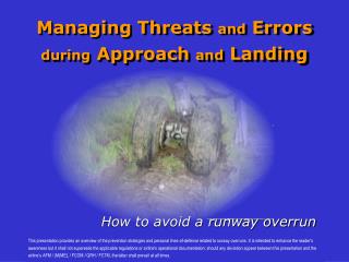
Mechanics and control of the pes planus versus normal foot during jumping and landing. Reporter: Reportor : Zong-Shein Chen Supervisor : Sai-Wei Yang. Introduction. Pes planus (flat foot) - the medial longitudinal arch of foot is lower than established normal parameters

E N D
Mechanics and control of the pes planus versus normal foot during jumping and landing Reporter: Reportor : Zong-Shein Chen Supervisor : Sai-Wei Yang
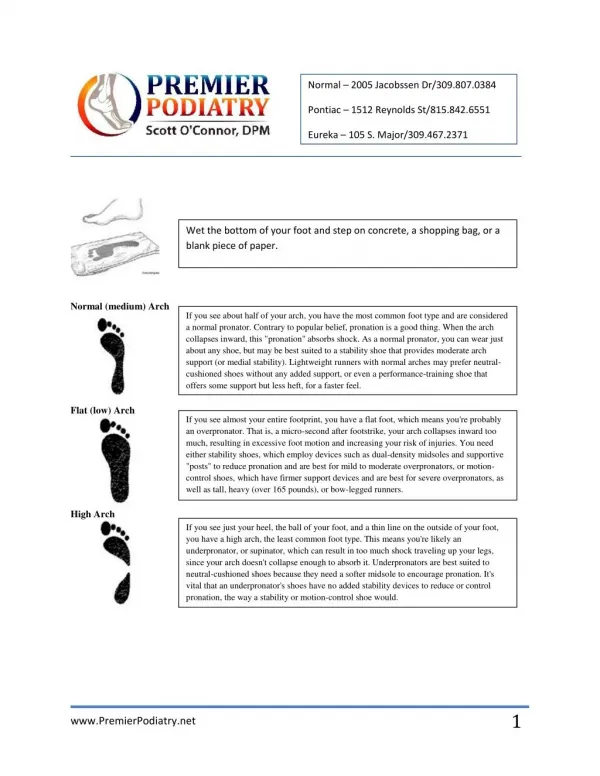
Introduction • Pes planus (flat foot) - the medial longitudinal arch of foot is lower than established normal parameters ~ Forrester D et al. Imaging of the foot and ankle;1988 - Occurs in approximately 15% of the population ~ Harris R and Beath T. J Bone Joint Surg;1948
The terms of pes planus : - Flexible : An observable medial arch during nonweightbearing and a flattening of the arch during weightbearing - Rigid : A stiff, flattened arch on and off weightbearing ~ Lee MS et al. J Foot Ankle Surg; 2005
The stages of pes planus : - In clinically and functionally, the rearfoot of flat foot subjects is valgus and the forefoot is varus ~ Bertani A et al. Clin Biomech. 1999 ~ Magee DJ. Orthopedic Physical Assessment; 2002
Abnormal biomechanical behaviors • The MLA collapses right down • Most of the plantar surface of foot contact with the ground • More strain on the plantar aponeurosis • Facilitating dorsiflexion • Unlocking of the midtarsal joint ~ Prost WJ. Fam Physician; 1979
Greater inversion ankle moment • Greater peak plantarflexion ankle moment • Less forefoot adduction • Less forefoot total transverse plane ROM ~ Hunt AE et al. Clin Biomech; 2004
Greater foot pressure under the second and third metatarsal heads • Greater foot pressure under the subhallucal area ~ Hunt GC. Examination of lower-extremity dysfunction; 1990
Injuries relate to pes planus • Pes planus are associated with a higher risk of injury among physically active people ~ Kulthanan T et al. J Med Assoc Thai; 2004 • Subjects with pes planus feet exhibited greater incidences of soft tissue and medial foot injuries and knee injuries ~ Williams DS et al. Clin Biomech; 2001
Sesamoiditis • Plantar fasciitis • Achilles tendinitis • Medial shin pain • Patello-femoral joint pain • Metatarsal stress fractures • Navicular and fibular stress fractures ~ Hunt AE et al. Clin Biomech; 2004
Posterior tibialis tendon dysfunction - Painful pes planus can often be associated with Posterior Tibial Tendon Dysfucntion (PTTD) - The posterior tibial muscle has a significant role in supporting the medial longitudinal arch ~ Kulig K et al. Med Sci Sports Exerc; 2005
Risk sports for foot and ankle injury ~ DeLee et al. Br J Sports Med; 2003
Jumping and landing • Jump-landing protocols have been used to measure postural sway • In an attempt to reduce the landing force the body must anticipate the landing and prepare for it by increasing muscle stiffness ~ McKinely P, Pedotti A. Exp Brain Res ;1992 • Further reduction of landing force can be accomplished by allowing the knee and hip to flex more which increases the time of landing providing an attenuation in kinetic energy ~ McNair P et al. Br J Sports Med; 2000
Effect of Foot Orthotics on Quadriceps and Gluteus Medius Electromyographic Activity During Selected Exercises ~ Hertel J, Sloss BR et al. Arch Phys Med Rehabil. 2005;86:26-30 • Design:Experimental, controlled • Participants: Thirty healthy young adults, 10 with each foot type- Foot type was subjectively categorized by a clinician experienced in lower extremity biomechanic evaluation
Interventions- 3 foot-type groups : pes planus, pes cavus, pes rectus- Each tested in 4 orthotic conditions : no orthotic, 7° medial rearfoot post, 4° lateral rearfoot post, and neutral rearfoot post- Performing 3 different exercises : single-leg squatting, lateral stepdown, and maximum vertical jump
Outcome Measure :Surface EMG activity for the vastus medialis, vastus lateralis, and gluteus medius during exercises • Results : Less vastus lateralis activity was found with the vertical jump with all orthotic conditions, regardless of foot type
Purpose • Few investigations focus on the jumping and landing biomechanical behaviors of pes planus subjects • The purpose in this study is to explore the jumping and landing biomechanical behaviors of pes planus subjects
Hypothesis • The jumping and landing biomechanical behaviors are different between pes planus and normal foot subjects, including COP excursion way, magnitude and direction of GRF, relative motion of foot-leg-knee and the EMG activity of the muscle
Methods - Participants • Thirty adult, aged from 18 to 25 years old • Inclusion criteria : - arch index > 0. 26 - flexible pes planus ~Williams DS et al. 2000
Exclusion criteria : - Acute foot injuries - Previous osseous foot surgery - Diagnosed with inflammatory arthritis, diabetes mellitus, congenital defects or neuromuscular disease
Instrumentation • Vision motion analysis system ~ VICON : to collect kinematic data • AMTI force plate : to collect kinetic data • Surface EMG : to collect muscle activation data, tibialis posterior, peroneus brevis and longus, medial and lateral gastrocnemius, vastus medialis and lateralis, biceps femoris
Procedures • While jumping, subjects were instructed to hold their hands at their waist to restrict arm movement substitution for jump effort • Four conditions, and 3 trials for each condition : 1. Vertical jump with both legs at maximum effort and to land with both legs 2. Vertical jump with both legs at maximum effort and to land on the dominant leg 3. Forward jump with both legs and to land with both legs 4. Forward jump with both legs and to land on the dominant leg
Data analysis • Descriptive statistics and two-way ANOVA tests will be used to compare the difference of testing order effects and four experimental conditions • Tukey’s post-hoctest will be used when an overall significant differencesare found • An alpha level of 0.05 will be used to test for significance
Reference • Lee MS, Vanore JV et al. Diagnosis and Treatment of Pediatric Flatfoot. J Foot Ankle Surg. 2005;43:341-373 • Hunt AE, Smith RM. Mechanics and control of the flat versus normal foot during the stance phase of walking. Clin Biomech. 2004;19:391-397 • Ledoux WR, Hillstrom HJ. The distributed plantar vertical force of neutrally aligned and pes planus feet. Gait posture. 2002;15:1-9 • Prost WJ. Biomechanics of the foot. Fam Physician. 1979;25:821-31 • Hunt GC. Orthopaedic and sports physical therapy. Examination of lower-extremity dysfunction. Second Edition. 1990:395-421 • Williams DS, McClay IS et al. Arch structure and injury patterns in runners. Clin Biomech. 2001;16:341-347 • Kulig K, Burnfield JM et al. Effect of foot orthoses on tibialis posterior activation on persons with pes planus. Med Sci Sports Exerc;2005:24-29 • Hertel J, Sloss BR et al. Effect of Foot Orthotics on Quadriceps and Gluteus Medius Electromyographic Activity During Selected Exercises. Arch Phys Med Rehabil. 2005;86:26-30