Download
1 / 4
60 likes | 614 Vues
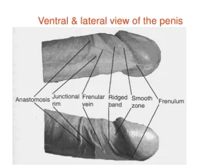
Ventral & lateral view of the penis. Evolution of foreskin ( prepuce).

E N D
Like the scrotal dartos muscle, the prepuce muscle is composed of unstriped fibers which form an incomplete investment of the penis from its base to the extremity of the prepuce. The muscle is situated 1 or 2 mm beneath the skin .The muscle fibers are very slender & are made of a few muscle cells only, but they run in every direction: transversely, longitudinally or obliquely. The fibers are loosely packed. The area between muscle cells is filled up with connective tissue. There is of course no subcutaneous tissue Peripenic muscle
The peripenic muscles are oriented in specific way in the prepuce. The muscle bundles are radially oriented to form a kind of sphincter which tend to close the orifice when the penis is flaccid. In pathological condition , it forms phimosis which can cause back flow of the urinary stream. Moreover, all extraneous matter ( including urine, sand,..) is prevented from gaining access to the preputal space. Phimosis