Download

1 / 25
250 likes | 500 Vues
Prenatal Evaluation of High Risk Pregnancies following Invitro -Fertilization: “Reduce Mother and Fetus Mortality Rates!”. Irani Sh. * ( B.Sc.&PHD ), Javam M. ( B.Sc ), Ahmadi F. (MD )

E N D
Prenatal Evaluation of High Risk Pregnancies following Invitro-Fertilization: “Reduce Mother and Fetus Mortality Rates!” Irani Sh.* (B.Sc.&PHD), Javam M. (B.Sc), Ahmadi F. (MD) Department of Reproductive Imaging at Reproductive Biomedicine Research Center, Royan Institute for Reproductive Biomedicine, ACECR, Tehran, Iran
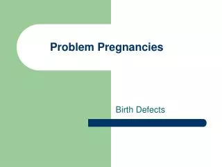
What’s the issue ? • The use of assisted-reproduction technologies has increased over the past decade! • Pregnancies following IVF are associated with higher risk of obstetric morbidities and perinatal mortality and several studies have demonstrated that the rate of prenatal complications is significantly more frequent in IVF-conceived pregnancies compared to spontaneous pregnancies.
Thus, a proper prenatal evaluation is required in this group to protect mother and fetus health! • Sonographicexamination during pregnancy is a helping method to detect pregnancy complications and to organize a proper “prenatal care” for IVF-conceived women. • We provide a clinical instruction for sonographic assessment of pregnancies following IVF and management of patients based on reports:
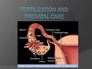
1st trimester : • Goals of first ultrasound scan of IVF-resulted pregnancies: • To assure a normal “intrauterine” pregnancy! • To rule out emergency conditions that are a threat to mother’s health: • * Ectopic or Heterotopic Pregnancy is shown to be 5-10 times more prevalent • than general population! • * Molar pregnancy& threatened abortion are also more frequent in this group • To establish gestational age “accurately” and to confirm fetus viability
To determine the number of fetuses and chorionicity-amnionicity in multiple pregnancies based on: • the number of gestational sacs, amnions, and yolk sacs M-D twins (MZ) M-M twins (MZ) D-D twins (DZ)
1st trimester complications: 1. Bleeding • without IUP: • with IUP: • Complete Abortion • Very early pregnancy • Retained product of P. • Tubal ectopic • Complete hydatyformMole (less common) • Abdominal ectopic (rare) • Failed pregnancy (missed AB FHR -) • Hemorrhage • Partial mole (less common) • An embryonic • Heterotopic pregnancy (rare) • Interestitial / cervical EP (rare) • Twin demise
2. Pain: • Hemorrhagic cyst • Corpus Luteumcyst • EP • OHSS • Adnexal torsion • Appendicitis (less common)
3. Discrepancy between GA and ET-date: • GS without fetal pole • Fetal pole without FHR • Fetal pole with FHR • Multiple fetuses blighted ovum or not ? (GS diameter <20mm or >20mm?) monitor sonography recommended (based on previous exams) follow-upwithin weeks 11-14 follow-up studies based on chorionicity
1st trimester Anomaly Screening • Some embriologists believe that the risk of fetal anomalies in IVF-resulted embryos is a bit higher! • Thus, a careful examination needs to be done for anomaly screening in this group. • First anomaly scan contains “Nuchal Translucency” measurement and look for structural abnormalities within 11th-14th weeks. • Further investigations are recommended in case of increased risk.
First Trimester Anomaly Screening
2nd & 3rd trimesters : • Second and third trimester have are vital periods of fetal growth. • There are several complications which have adverse affects on this event. • Sonographyis helpful for early detection of risks for most of these complications such as vaginal bleeding, IUGR, preterm birth, etc.
1. Cervical Insufficiency: • A prevalent cause of preterm birth • among these women • Early diagnosis & decision for cerclage • placement can reduce PTB rate. • Screening method: • - Serial cervical length measurement by means of TAS or TVS (preferred) • - At least twice during 2nd trimester (before 20th week)
2. Bleeding: • Common reasons: • Placenta previa • Marginal sinus previa • Placenta accreta spectrum • Less common: • Succenturiate lobe • Placenta accreta • Placenta abruption • Cesarean section rupture Marginal previa
3. PROM & oligohydroamnios: • Maximum Vertical pocket<2cm , AFI < 5cm • Early oligohydroamnios: <25 week • Prolonged oligohydroamnios : >14 days • D.D: PROM / IUGR /Renal agenesis /bladder outlet obstruction / TTTS in multiples • Serial monitoring sonographies are essential
4. Growth Restriction: • Fetal weight < %10 percentile • IUGR differential diagnosis approach: • Rule out fetal anomaly as cause for IUGR • Amniocentesis if fetal anomaly suspected • Consider maternal medical history • Assess amniotic fluid • Assess fetal / placental circulation • Differentiate between symmetric vs. asymmetric IUGR • Doppler Examination • Biophysical profile (BPP) if needed
5. Gestational Diabetes and Pre-eclampsia: - Consider fetal growth and probable discrepancy - Mention to placenta size and shape - Assess A.F.I - Use Color Doppler to check blood circulation if needed - BPP if prescribed
Discrepancy between multiples: • “Consider Chorionicity!” • Follow up sonography every 2 weeks
Recommendation for US exams: 1. Early first trimester ( to week of gestation): To confirm intra uterine pregnancy (IUP) and to rule out ectopic pregnancy (EP), to ensure fetal heart activity, diagnosis of multiple pregnancy and estimation of gestational age. 2. Late first trimester (to week): To evaluation fetal growth, to assess fetal structural malformations (like Anencephaly, etc.) and detection of anomaly markers (measuring nuchal translucency) to find out high risk patients. 3. Second trimester ( to week): Evaluation of fetal growth, to assess fetal structural malformations and anomaly markers (measuring nuchal fold & nasal bone) to detect high risk patients, diagnosis of cervical insufficiency and need to “cerclage placement”. 4. Third trimester ( to week): Evaluation of fetal growth and weight, measuring amniotic fluid index (AFI), assessment of fetal structural abnormalities (such as hydrocephaly, etc.) and Biophysical Profile (if needed).
Conclusion: • Prenatal sonography is an accurate, non-invasive, and cost-effective tool that helps midwives and obstetricians to evaluate mother and fetus health and detection of pregnancy complications to give better prenatal care for pregnant women, especially for those who became pregnant after IVF treatment. • A proper “prenatal care” can be organized based on sonography reports.
Thanks for your Attention!