Download
1 / 29
300 likes | 611 Vues
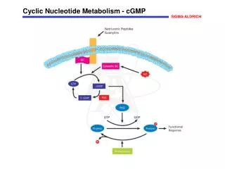
cGMP Intracellular Signal. cGMP is made from GTP by the enzyme gaunylyl cyclase . Atrial natriuretic peptide and nitric oxide function through this Signal. These are potent vasodilators. Inhibitors of cGMP phosphodiestrase is sildenafil (Viagra).

E N D
cGMP Intracellular Signal • cGMP is made from GTP by the enzyme gaunylylcyclase. • Atrialnatriuretic peptide and nitric oxide function through this Signal. • These are potent vasodilators. • Inhibitors of cGMPphosphodiestrase is sildenafil (Viagra).
The increased cGMP activates cGMP dependent protein kinase (PKG). • This in turn phosphorylates a number of smooth muscle proteins. • This leads to relaxation of smooth muscle cell and vasodilation.
Calcium or Phosphatidylinositol • Ionized calcium is an important regulator of many cellular processes, including: • muscle contraction, • secretion, • blood clotting, • enzyme activity and • membrane excitability.
Calcium Metabolism: • Extracellular Ca conc is 5mmol/L. • Ca is restrained from entering the cell and the intracellular conc of free and ionized Ca is very low 0.05-10μmol/L. • Some signal must provide communication between the hormone receptor on the plasma membrane and the intracellular Ca reservoirs.
This is accomplished by products of phosphatidylinositolmetabolism. • Cell surface receptors such as those for: • acetylcholine, • antidiuretic hormone and • α1 – catecholamines • When occupied by these ligands they are potent activators of Phospholipase C.
This involves a specific G protein, which also may activate a calcium channel. • Phospholipase C cleaves Phosphatidylinositol 4,5 bisphosphate (PIP2) into : • 1. 1,2 Diacylglycerol(DAG) and • 2. inositol 1,4, 5 triphosphate(IP3).
DAG is a potent activator of protein kinase C. • The activated PKC phosphorylates specific substrates, which then alter physiologic processes.
IP3 liberates stored intracellular Ca from the endoplasmic reticulum. • Ca-Calmodulin complex is formed and this also activate specific kinases which phosphorylates specific substrates, which then alter physiologic processes. • The same G- protein activation also activates a calcium channel and Ca can enter the cell.
Enzymes and Proteins regulated by Ca and Calmodulin • AdenylylCyclase • Ca –dependent protein kinases. • Ca- Mg ATPase • Nitric oxide synthase • Phosphorylase kinase
Insulin Signaling Pathways • Insulin is released in response to hyperglycemia. • Insulin binds to cell surface receptors . • These receptors have intrinsic tyrosine kinase activity. • The receptors are then auto phosphorylated on tyrosine residues. • This initiates a complex series of events.
The phosphorylated receptor then next phosphorylate insulin receptor substrates(IRS 1-4). • These IRS then bind to Src homology domains on the proteins and are involved in different effects of insulin.
Effects of this pathway: • 1. Protein translocation (glucose transporters, insulin receptors) • Enzyme activity (insulin receptor, Phosphatases, phosphodiestrases) • Gene transcription (PEPCK, Glucagon,Glucokinase)
JAK STAT Pathway • Growth hormone • Prolactin • Erythropoietin • Cytokines • Activate a tyrosine kinase, but this activity is not an integral part of the receptor.
When the ligand binds to the receptor then the receptor dimerizes and associated cytoplasmic protein kinases such as; Tyk-2, Jak1, Jak2 are phosphorylated. • Jak-P now becomes an active kinase and it then phosphorylates the receptor on tyrosine residue.
The kinases then phosphorylate other cytoplasmic proteins. • One of the cytoplasmic proteins family is called signal transducers and activators of transcription(STAT).
The phosphorylated STAT protein dimerizes and translocates to the nucleus and bind to specific DNA sequence. • The phosphotyrosine residues of the receptor also bind to docking proteins through SH2 • domain. • This result in the activation of other pathways.
NF-кB Pathway • This pathway is regulated by Glucocorticoids. • NF-кB factor is composed of two subunits termed p50 and p65. • Normally NF-кB factor is sequestered in the cytoplasm in an inactive form by the inhibitors(IкB).
Extracellular stimuli such as proinflammatory cytokines, reactive oxygen species, and mitogens lead to the activation of IкB kinase complex, called IKK. • IKK phosphorylate inhibitor(IкB). • This causes degradation of inhibitor(IкB). • NF-кB factor is free and it translocates to the nucleus and promote gene transcription.
NF-кB activators are: • Proinflammatory cytokines • Bacterial and viral infection • Reactive oxygen species • Mitogens
Inhibitors of NF-кB: • Glucocorticoid hormones are therapeutically useful agents for the treatment of a variety of inflammatory and immune diseases.. • These actions in part are explained by the inhibition of NF-кB pathway. • Increase the level of inhibitoe • Compete with the Co activators for the receptor. • Directly bind to p65 subunit of NF-kB.