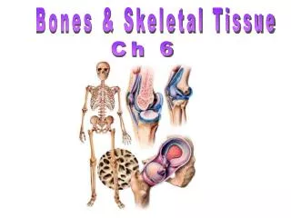
Chapter 6 The Skeletal System:Bone Tissue
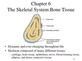
Chapter 6 The Skeletal System:Bone Tissue. Dynamic and ever-changing throughout life Skeleton composed of many different tissues cartilage, bone tissue, epithelium, nerve, blood forming tissue, adipose, and dense connective tissue. Functions of Bone. Supporting & protecting soft tissues

Chapter 6 The Skeletal System:Bone Tissue
E N D
Presentation Transcript
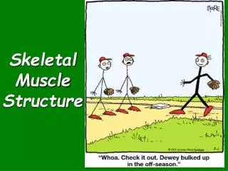
Chapter 6The Skeletal System:Bone Tissue • Dynamic and ever-changing throughout life • Skeleton composed of many different tissues • cartilage, bone tissue, epithelium, nerve, blood forming tissue, adipose, and dense connective tissue
Functions of Bone • Supporting & protecting soft tissues • Attachment site for muscles making movement possible • Storage of the minerals, calcium & phosphate -- mineral homeostasis • Blood cell production occurs in red bone marrow (hemopoiesis) • Energy storage in yellow bone marrow
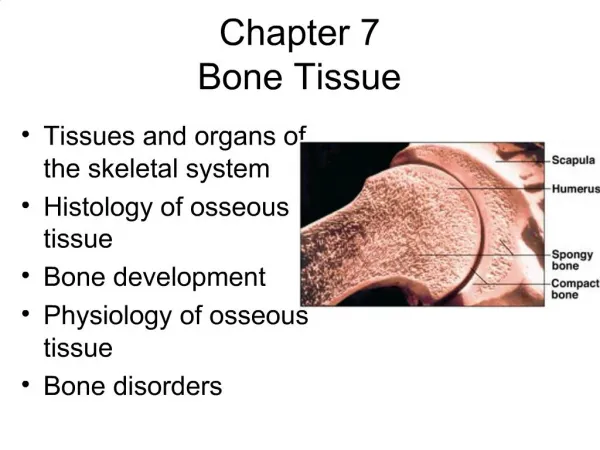
Anatomy of a Long Bone • Diaphysis = shaft • Epiphysis = one end of a long bone • Metaphysis = growth plate region • Articular cartilage over joint surfaces acts as friction & shock absorber • Medullary cavity = marrow cavity • Endosteum = lining of marrow cavity • Periosteum = tough membrane covering bone but not the cartilage • fibrous layer = dense irregular CT • osteogenic layer = bone cells & blood vessels that nourish or help with repairs
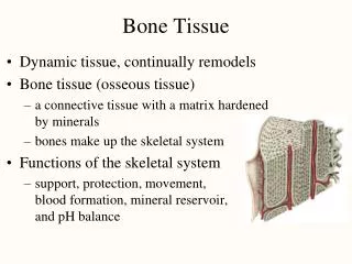
Histology of Bone • A type of connective tissue as seen by widely spaced cells separated by matrix • Matrix of 25% water, 25% collagen fibers & 50% crystalized mineral salts • 4 types of cells in bone tissue
Cell Types of Bone • Osteoprogenitor cells ---- undifferentiated cells • can divide to replace themselves & can become osteoblasts • found in inner layer of periosteum and endosteum • Osteoblasts--form matrix & collagen fibers but can’t divide • Osteocytes ---mature cells that no longer secrete matrix • Osteoclasts---- huge cells from fused monocytes (WBC) • function in bone resorption at surfaces such as endosteum
Matrix of Bone • Inorganic mineral salts provide bone’s hardness • hydroxyapatite (calcium phosphate) & calcium carbonate • Organic collagen fibers provide bone’s flexibility • their tensile strength resists being stretched or torn • Mineralization (calcification) is hardening of tissue when mineral crystals deposit around collagen fibers • remove minerals with acid & rubbery structure results • Bone is not completely solid since it has small spaces for vessels and red bone marrow • spongy bone has many such spaces • compact bone has very few
Compact or Dense Bone • Looks like solid hard layer of bone • Makes up the shaft of long bones and the external layer of all bones • Resists stresses produced by weight and movement
Histology of Compact Bone • Osteon is concentric rings (lamellae) of calcified matrix surrounding a vertically oriented blood vessel • Osteocytes found in spaces called lacunae • Osteocytes communicate through canaliculi filled with extracellular fluid that connect one cell to the next cell • Interstitial lamellae represent older osteons that have been partially removed during tissue remodeling
The Trabeculae of Spongy Bone • Latticework of thin plates of bone called trabeculae oriented along lines of stress • Spaces in between these struts are filled with red marrow where blood cells develop • Found in ends of long bones and inside flat bones such as the hipbones, sternum, sides of skull, and ribs. No true Osteons.
Blood and Nerve Supply of Bone • Periosteal arteries • supply periosteum • Nutrient arteries • enter through nutrient foramen • supplies compact bone of diaphysis & red marrow • Metaphyseal & epiphyseal aa. • supply red marrow & bone tissue of epiphyses
Bone Formation or Ossification • All embryonic connective tissue begins as mesenchyme. • Intramembranous bone formation = formation of bone directly from mesenchymal cells. • Endochondral ossification = formation of bone within hyaline cartilage.
Bone Growth in Length • Epiphyseal plate or cartilage growth plate • cartilage cells are produced by mitosis on epiphyseal side of plate • cartilage cells are destroyed and replaced by bone on diaphyseal side of plate • Between ages 18 to 25, epiphyseal plates close. • cartilage cells stop dividing and bone replaces the cartilage (epiphyseal line) • Growth in length stops at age 25
Zones of Growth in Epiphyseal Plate • Zone of resting cartilage • anchors growth plate to bone • Zone of proliferating cartilage • rapid cell division (stacked coins) • Zone of hypertrophic cartilage • cells enlarged & remain in columns • Zone of calcified cartilage • thin zone, cells mostly dead since matrix calcified • osteoclasts removing matrix • osteoblasts & capillaries move in to create bone over calcified cartilage
Bone Growth in Width • Only by appositional growth at the bone’s surface • Periosteal cells differentiate into osteoblasts and form bony ridges and then a tunnel around periosteal blood vessel. • Concentric lamellae fill in the tunnel to form an osteon.
Bone Remodeling • Ongoing since osteoclasts carve out small tunnels and osteoblasts rebuild osteons. • osteoclasts form leak-proof seal around cell edges • secrete enzymes and acids beneath themselves • release calcium and phosphorus into interstitial fluid • osteoblasts take over bone rebuilding • Continual redistribution of bone matrix along lines of mechanical stress • distal femur is fully remodeled every 4 months
Fracture is break in a bone Healing is faster in bone than in cartilage due to lack of blood vessels in cartilage Healing of bone is still slow process due to vessel damage Clinical treatment closed reduction = restore pieces to normal position by manipulation open reduction = surgery Fracture & Repair of Bone
Calcium Homeostasis & Bone Tissue • Skeleton is reservoir of Calcium & Phosphate • Calcium ions involved with many body systems • nerve & muscle cell function • blood clotting • enzyme function in many biochemical reactions • Small changes in blood levels of Ca+2 can be deadly (plasma level maintained 9-11mg/100mL) • cardiac arrest if too high • respiratory arrest if too low
Pull on bone by skeletal muscle and gravity is mechanical stress . Stress increases deposition of mineral salts & production of collagen (calcitonin prevents bone loss) Lack of mechanical stress results in bone loss reduced activity while in a cast astronauts in weightlessness bedridden person Weight-bearing exercises build bone mass (walking or weight-lifting) Exercise & Bone Tissue
Osteoporosis • Decreased bone mass resulting in porous bones • Those at risk • white, thin menopausal, smoking, drinking female with family history • athletes who are not menstruating due to decreased body fat & decreased estrogen levels • people allergic to milk or with eating disorders whose intake of calcium is too low • Prevention or decrease in severity • adequate diet, weight-bearing exercise, & estrogen replacement therapy (for menopausal women) • behavior when young may be most important factor