Download
1 / 45
450 likes | 719 Vues
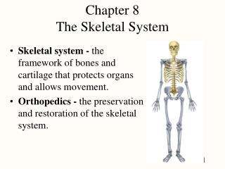
Chapter 5 The Skeletal System Provides an internal framework for the body, protects organs by enclosure, and anchors skeletal muscles so that muscle contractions can cause movement. Bone Formation, Growth, & Remodeling. Types of Bone Cells. Osteocytes Mature bone cells Osteoblasts

E N D
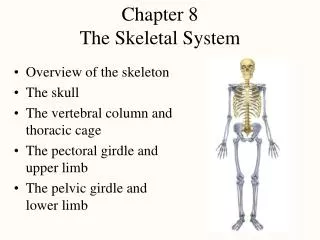
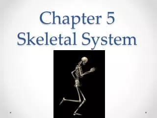
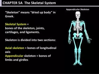
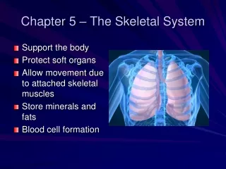
Chapter 5The Skeletal System Provides an internal framework for the body, protects organs by enclosure, and anchors skeletal muscles so that muscle contractions can cause movement
Types of Bone Cells • Osteocytes • Mature bone cells • Osteoblasts • Bone-forming cells • Osteoclasts • Bone-destroying cells • Break down bone matrix for remodeling and release of calcium • Bone remodeling is a process performed by both osteoblasts and osteoclasts
Changes in the Human Skeleton • In embryos, the skeleton is primarily hyaline cartilage • During development, much of this cartilage is replaced by bone • Cartilage remains in isolated areas • Bridge of the nose • Parts of ribs • Joints
Bone Growth • Epiphyseal plates allow for growth of long bones during childhood • New cartilage is continuously formed • Older cartilage becomes ossified • Cartilage is broken down • Bone replaces cartilage
Bone Remodeling • Bones are remodeled continually in response to changes in 2 factors • Calcium levels in the blood • Pull of gravity & muscles on the skeleton
Rickets • Softening & weakening of bones in children • Due extreme & prolonged vitamin D deficiency • Vitamin D is essential in promoting absorption of calcium & phosphorus from the GI tract Required to build strong bones • Adding vitamin D and calcium to the diet Corrects any resulting bone problems • Some skeletal deformities Surgery • Symptoms • Skeletal deformities • Bowed legs, spine curvature, pelvic deformities, breast bone projection • Fragile bones, impaired growth in height, dental problems, bone pain, muscle weakness
OsteoporosiswebsiteERT websiteMenopause site • Disease of the bones: • Thin, brittle bones with lots of holes • Bones are susceptible to fractures • Especially, the hips, spine, & wrists • Usually strikes after age 60 • Symptoms: • Broken bone after a fall • Back pain • Decreased height • Curved backbone • Cause: • Lack of bone strength or bone density
Osteoporosis • Risk Factors: • Age • Female gender • Menopause • After menopause Reduced production of estrogen, which protects the body from bone loss • Family history – Genetics • Slender body frame • Race European; Asian • Smoking • Lack of weight-bearing exercise • Alcohol • Lack of calcium & vitamin D in the diet • Osteoporosis Slideshow
Osteoporosis • Treatment: • Medication • Fosamax: Reduces bone loss & build bone thickness • Calcium & Vitamin D supplements • Diet • Eat dark green vegetables, yogurt, milk Calcium • Eat eggs, fatty fish, fortified cereal Vitamin D • Exercise Weight bearing
Bone Fractures • A break in a bone • Types of bone fractures • Closed (simple) fracture – break that does not penetrate the skin • Open (compound) fracture – broken bone penetrates through the skin • Bone fractures are treated by reduction and immobilization • Realignment of the bone
Common Types of Fractures * Know Table 5.2 page 137
Repair of Bone Fractures Hematoma (blood-filled swelling) is formed Break is splinted by fibrocartilage to form a callus Fibrocartilage callus is replaced by a bony callus Bony callus is remodeled to form a permanent patch
Stages in the Healing of a Bone Fracture * KNOW Figure 5.5 page 138
Joints • Articulations of bones • Functions of joints • Hold bones together • Allow for mobility • Joints are classified in 2 ways • Functionally • Structurally
Functional Classification of Joints • Focuses on the amount of movement allowed by the joint • Synarthroses – Immovable joints • Amphiarthroses – Slightly moveable joints • Immovable & slightly movable joints Restricted to the axial skeleton • Firm attachments & protection of internal organs • Diarthroses – Freely moveable joints • Predominantly in the limbs
Structural Classification of Joints • Fibrous joints • Generally immovable • Cartilaginous joints • Immovable or slightly moveable • Synovial joints • Freely moveable
Fibrous Joints • Bones united by fibrous tissue • Ex. Sutures of the skull • Synarthroses (largely immovable)
Cartilaginous Joints • Bone ends are connected by cartilage • Examples • Amphiarthrotic- Slightly movable • Pubic symphysis • Intervertebral joints • Synarthrotic • Immovable • True ribs & sternum
Synovial Joints • Articulating bones are separated by a joint cavity • Synovial fluid is found in the joint cavity
Features of Synovial Joints- Diarthroses • Articular (hyaline) cartilage) covers the ends of bones • Joint surfaces are enclosed by a fibrous articular capsule • Have a joint cavity filled with synovial fluid • Ligaments reinforce the joint
Structures Associated with the Synovial Joint • Bursae – flattened fibrous sacs • Lined with synovial membranes • Filled with synovial fluid • Not actually part of the joint • Common where ligaments, muscles, skin, tendons, or bones rub together • Tendon sheath • Elongated bursa that wraps around a tendon subject to friction
Types of Synovial Joints: Based on Shape
Inflammatory Conditions Associated with Joints • Bursitis – inflammation of a bursa usually caused by a blow or friction • Tendonitis – inflammation of tendon sheaths • Arthritis – inflammatory or degenerative diseases of joints • Over 100 different types • The most widespread crippling disease in the United States
Clinical Forms of Arthritis • Osteoarthritis • Most common form of chronic • arthritis • Chronic degenerative condition • Probably related to the normal aging • processes • “Wear-and-tear arthritis” • Affects the articular cartilages • Most commonly affected joints • Fingers, cervical & lumbar regions of the spine • Large weight-bearing joints of the lower limbs (knees & hips) • Progression = slow & irreversible, but rarely crippling
Clinical Forms of Arthritis • Rheumatoid arthritis • Chronic inflammatory disorder • Autoimmune disease- Disorder in which the body’s immune system attempts to destroy its own tissues • Initial trigger = unknown • Usually occurs between the ages of 40 and 50 but it may occur at any age • Affects 3 times as many women as men • Joints of the fingers, wrists, ankles, and feet are affected at the same time and usually in a symmetrical manner • Often leads to deformities
Clinical Forms of Arthritis • Gouty Arthritis “Gout” • Disease in which uric acid accumulates in the blood and may be deposited as needle-shaped crystals in the soft tissues of joints • Agonizing painful attack- typically affects the great toe • Most common in males
TMJ continued… • For me the connection is fairly obvious when viewing radiological images of people with atlas subluxations it becomes patently obvious that the jaw mandible and hence the TMJ are out of alignment. The crooked or tilted head (X-ray opposite) sitting atop the cervical spine results in non-alignment or disarticulation of the TMJ in the cranial fossa (recess). The joints do not work properly, with the disc being captured during opening and/or closing, and the neck and shoulder muscles going into painful spasm during the normal process of eating.