Chapter 6 The Skeletal System
Summary. StructureFunctionsGrowthEmbryonicFactors that affect bone growth and maintenanceTypes of bonesDivisions of the skeletonJoints/articulations. Structure. There are a total of 206 bones in the human body.The skeletal system contains both bone and cartilage

Chapter 6 The Skeletal System
E N D
Presentation Transcript
1. Chapter 6 The Skeletal System Bones and Cartilage
2. Summary Structure
Functions
Growth
Embryonic
Factors that affect bone growth and maintenance
Types of bones
Divisions of the skeleton
Joints/articulations
3. Structure
There are a total of 206 bones in the human body.
The skeletal system contains both bone and cartilage � 2 types of connective tissue.
The appearance and texture of bone varies, depending on its location.
4. Structure Compact Bone
Outer layer of bone, very hard and dense.
Organized in structural units called Haversian systems.
Matrix is composed of Ca salts (Ca carbonate and Ca phosphate)
Osteocytes � living bone cells that live in matrix.
5. Structure Porous (Spongy) bone
Located in the ends of long bones.
Many spaces that are filled with red bone marrow which produces blood cells.
Trabeculae � needle-like threads of spongy bone that surround the spaces. Add strength to this portion of the bone.
7. Structure Cartilage
Matrix is a firm gel with chondrocytes suspended in the matrix.
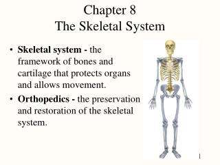
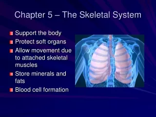
8. Functions Provides framework that supports the body.
Protection
Provides movement with the help of the skeletal muscles.
**Storage of calcium
Produces blood cells
9. Embryonic Bone Growth The skeleton is first made of cartilage and fibrous connective tissue. These are gradually replaced by bone.
Bone matrix is produced by osteoblasts.
�Calcification� begins at 8 weeks gestation.
Process is not complete at birth.
12. Factors that Affect Bone Growth and Maintenance Heredity �
Nutrition �
13. Factors that Affect Bone Growth and Maintenance Hormones � growth hormone, thyroxine, parathyroid hormone and insulin help regulate cell division, protein synthesis, Ca metabolism and energy production.
Exercise or �stress� � �bearing weight� causes bones to thicken.
14. 4 Types of Bones Long bones � bones of the arms and legs. Structure:
Diaphysis �
Medullary cavity �
15. 4 Types of Bones Long bones cont�d
Epiphysis
Composed of spongy bone which contains red bone marrow in children, replaced with yellow marrow in adults.
Epiphyses are covered with compact bone which in turn is covered with articular (hyaline) cartilage.
Periosteum �
Endosteum �
17. 4 Types of Bones Short bones � carpels and tarsals (wrist and ankle).
Flat bones � skull bones.
Irregular bones � vertebrae.
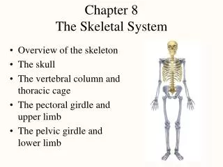
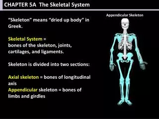
18. Divisions of the Skeleton Axial Skeleton � bones of the skull, spine, chest and the hyoid bone.
Skull � 8 bones that form the cranium, 14 that form the face and 6 in the middle ears.
Frontal � forehead bone, also forms upper part of eye sockets.
Parietal � top/upper sides of the cranium behind the frontal bone
Temporal � forms the lower sides of the cranium; contains middle and inner ear structures, external auditory canal, and mastoid process (behind the ear).
19. Divisions of the Skeleton Axial skull
Cranium
Occipital bone - forms the back of the skull, large hole (foramen magnum) allows entry of spinal cord.
Sphenoid � forms central part of the floor of the cranium; pituitary gland is located in a depression called the sella turcica (Turkish sadle).
Ethmoid bone � helps form floor of cranium, side walls and roof of nose.
Nasal bones form upper bridge of nose.
21. Divisions of the Skeleton Axial - skull
Face
Maxilla � upper jawbones
Zygomatic bones � cheek bones; help form eye orbit.
Mandible � lower jawbone.
Lacrimal bones � form medial wall of the eye socket and side of nasal cavity.
Palatine � forms back part of the roof of the mouth, part of the orbit.
Vomer � forms lower, back part of nasal septum.
24. Divisions of the Skeleton Axial - skull
Ear bones � from the outside in..
Malleus � hammer
Incus � anvil
Stapes � stirrup
Sinuses � spaces or cavities inside the cranium
Sutures � Immovable joints that join skull bones together
Lamboidal � between the parietal and occipital
Squamous � between the parietal and temporal
Coronal � between parietal and frontal
Sagittal � between parietal bones
**Fontanels � usually ossify by 2 years of age
26. Divisions of the Skeleton Vertebral column consists of a series of separate bones or vertebrae connected such that they form a flexible curved rod.
7 Cervical vertebrae - Atlas and axis are first two vertebrae
12 Thoracic
5 Lumbar
Sacrum
Coccyx
28. Divisions of the Skeletal System Axial � Vertebral column
Vertebral column has 4 curves that support the weight of the body and help with balance.
Cervical curvature � curves anteriorly
Thoracic curvature curve posteriorly
Lumbar curvature � anteriorly
Sacral � posteriorly
29. Divisions of the Skeletal System Axial � Vertebral column
Abnormal curves
Kyphosis (thoracic) = exagerated curve =
Lodosis (lumbar) = exagerated curve =
Scoliosis � lateral curve.
31. Divisions of the Skeletal System Axial � Thorax
Includes manubrium, sternum, thoracic vertebrae and the ribs.
All 12 ribs are all attached posteriorly to the vertebrae:
�True ribs� - 1st 7 pair are attached to the sternum by the costal cartilage beginning at the manubrium (upper part of the sternum).
False ribs � ribs 8, 9, and 10 are attached to the cartilage of the 7th ribs.
Floating ribs � last 2 pairs are not attached at the anterior end.
33. Divisions of the Skeletal System Appendicular skeleton � bones of the upper and lower extremities and their girdles.
Upper extremity
Scapula � shoulder blade
Clavicle � collar bone
Humerus � long bone of the upper arm; 2nd longest bone in the body. The humerus is attached to the scapula proximally and articulates with the radius and ulna distally to form the elbow joint.
34. Divisions of the Skeletal System Appendicular skeleton - Upper Extremity cont�d
Radius and ulna � bones of the lower arm, articulate with each other distally and with the carpals.
Carpals � 8 wrist bones
Metacarpals � palm of hand
Phalanges � 14 finger bones, 3 in each finger and 2 in each thumb.
36. Divisions of the Skeletal System Appendicular skeleton - Lower extremity
Pelvic (hip) girdle connects legs to the trunk. Consists of 2 large os coxae one on each side of the pelvis. Each os coxae consists of 3 separate bones in the infant:
Ilium
Ischium
Pubis These grow together in the adult.
37. Divisions of the Skeletal System Appendicular skeleton � Lower extremity
Femur � thigh bone, longest bone in the body. The femur fits into the cup-shaped �socket� = acetabulum � in the pelvic girdle. Distally the femur articulates with the patella (knee cap) and the�
Tibia (lower leg - shinbone).
Medial malleolus � inner anklebone, protuberance of the tibia.
Fibula � long slender, non-weight bearing bone located along the lateral border of the lower leg.
38. Divisions of the Skeletal System Appendicular skeleton � Lower extremity
Lateral Malleolus � rounded projection at the lower end of the fibula (outer anklebone)
Tarsals � 7 ankle bones; the talus articulates with the tibia and the calcaneous, the heel bone.
Metatarsals � 5 long bones of the foot.
Phalanges - toe bones, two in the �big� toe and 3 in each of the other toes.
41. Joints =Articulations Three types of joints � synarthroses, amphiarthroses and diarthroses
Synarthroses �
Amphiarthroses �
42. Joints =Articulations Diarthroses � freely moveable joints.
All have a joint capsule, a joint cavity and a layer of cartilage over the ends of the adjoining bones.
Joint Capsule � made of tough fibrous connective tissue and lined with the smooth slippery synovial membrane; fits over the ends of the bones like a sleeve.
Ligaments � cords or bands of fibrous connective tissue that hold the bones together firmly.
43. Joints =Articulations Diarthroses cont�d
Joint structure cont�d
Articular cartilage � layer of cartilage over the end of the bones.
Synovial membrane � secretes a lubricating fluid (synovial fluid) that allows easier movement with less friction.
44. Joints =Articulations Types of Diarthroses
Ball and Socket � ball like head on one bone fits into a socket of another. Ex � shoulder and hip. Widest range of movement.
Hinge � allow movements in only 2 directions � flexion and extension. Ex � elbow and knee, fingers.
Pivot � small projection of one bone pivots in an arch of another. Ex � atlas and axis.
45. Joints = Articulations Types of Diartroses
Saddle or saddle-shaped � allows for flexion, extension, abduction, adduction, opposition. **Only example � metacarpal bone of thumb and a carpal bone of the wrist.
Gliding � least moveable, flat articulating surfaces. Ex � carpals.
Condyloid � distal end of radius into the depressions in the carpals.