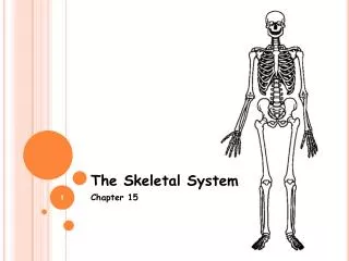
Chapter 6: Skeletal System I
Chapter 6: Skeletal System I. Bernard Siegfried Albinus 1697 – 1770 Famous for his drawings in the work entitled Tables of the Skeleton and Muscles of the Human Body published in 1747. An example of Albinus’ drawings of the skeleton.

Chapter 6: Skeletal System I
E N D
Presentation Transcript
Chapter 6: Skeletal System I
Bernard Siegfried Albinus 1697 – 1770 Famous for his drawings in the work entitled Tables of the Skeleton and Muscles of the Human Body published in 1747.
Figure 6.1 The bones and cartilages of the human skeleton. Epiglottis Larynx Thyroid cartilage Cartilage in external ear Cartilages in nose Trachea Cricoid cartilage Lung Articular Cartilage of a joint Cartilage in Intervertebraldisc Costal cartilage Respiratory tube cartilages in neck and thorax Bones of skeleton Pubic symphysis Axial skeleton Meniscus (padlike cartilage in knee joint) Appendicular skeleton Cartilages Articular cartilage of a joint Hyaline cartilages Elastic cartilages Fibrocartilages
Figure 6.2 Classification of bones on the basis of shape. (c) Flat bone (sternum) (a) Long bone (humerus) (b) Irregular bone (vertebra), right lateral view (d) Short bone (talus)
Figure 6.3a The structure of a long bone (humerus of arm). Articular cartilage Proximal epiphysis Spongy bone Epiphyseal line Periosteum Compact bone Medullary cavity (lined by endosteum) Diaphysis Distal epiphysis (a)
Figure 6.3b The structure of a long bone (humerus of arm). Articular cartilage Compact bone Spongy bone (b)
Figure 6.3c The structure of a long bone (humerus of arm). Endosteum Yellow bone marrow Compact bone Periosteum Perforating (Sharpey’s) fibers Nutrient arteries (c)
Figure 6.4 Comparison of different types of bone cells. (a) Osteogenic cell (b) Osteoblast (c) Osteocyte (d) Osteoclast Stem cell Matrix-synthesizing cell responsible for bone growth Mature bone cell that maintains the bone matrix Bone-resorbing cell
Figure 6.5 Flat bones consist of a layer of spongy bone sandwiched between two thin layers of compact bone. Spongy bone (diploë) Compact bone Trabeculae
Figure 6.9 Endochondral ossification in a long bone. Month 3 Birth Childhood to adolescence Week 9 Articular cartilage Secondary ossification center Spongy bone Epiphyseal blood vessel Area of deteriorating cartilage matrix Epiphyseal plate cartilage Hyaline cartilage Medullary cavity Spongy bone formation Bone collar Blood vessel of periosteal bud Primary ossification center 1 2 3 4 5 Cartilage in the center of the diaphysis calcifies and then develops cavities. Bone collar forms around hyaline cartilage model. The periosteal bud invades the internal cavities and spongy bone begins to form. The diaphysis elongates and a medullary cavity forms as ossification continues. Secondary ossification centers appear in the epiphyses in preparation for stage 5. The epiphyses ossify. When completed, hyaline cartilage remains only in the epiphyseal plates and articular cartilages.
Figure 6.11 Long bone growth and remodeling during youth. Bone remodeling Bone growth Articular cartilage Cartilage grows here. Epiphyseal plate Cartilage is replaced by bone here. Bone is resorbed here. Cartilage grows here. Bone is added by appositional growth here. Cartilage is replaced by bone here. Bone is resorbed here.
Figure 6.12 Parathyroid hormone (PTH) control of blood calcium levels. Calcium homeostasis of blood: 9–11 mg/100 ml BALANCE BALANCE Stimulus Falling blood Ca2+ levels Thyroid gland Osteoclasts degrade bone matrix and release Ca2+ into blood. Parathyroid glands Parathyroid glands release parathyroid hormone (PTH). PTH
Figure 6.13 Bone anatomy and bending stress. Load here (body weight) Head of femur Tension here Compression here Point of no stress
Figure 6.14 Vigorous exercise can lead to large increases in bone strength. (a) Cross- sectional dimension of the humerus Added bone matrix counteracts added stress (b) Serving arm Nonserving arm
Steel “Bone Cages” used to lengthen legs. These were originally developed in the Soviet Union in the 1950s to treat dwarfism.
Twelve-year-old boy with pituitary gigantism measuring 6'5" with his mother. Note the coarse facial features and prominent jaw.
Chelation Therapy – intravenous administration of chemicals designed to absorb toxic substances that have accumulated in the body. Most notably used for exposure to heavy metals such as lead or mercury.
Figure 6.15 Stages in the healing of a bone fracture. Hematoma External callus Bony callus of spongy bone Internal callus (fibrous tissue and cartilage) Healed fracture New blood vessels Spongy bone trabecula 1 2 3 4 A hematoma forms. Fibrocartilaginous callus forms. Bony callus forms. Bone remodeling occurs.
Figure 6.16 The contrasting architecture of normal versus osteporotic bone.
Figure 6.17 Fetal primary ossification centers at 12 weeks. Parietal bone Occipital bone Frontal bone of skull Mandible Clavicle Scapula Radius Ulna Ribs Humerus Vertebra Ilium Tibia Femur