Download
1 / 47
470 likes | 656 Vues
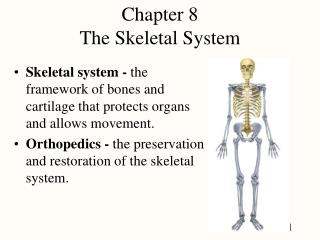
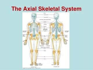
The Skeleton System Chapter 8/ Part I. Joe Pistack MS/ED. The Skeletal System. The Skeletal System. Consists of: Bones Joints Cartilage Ligaments The skeletal system consists of 206 bones. The Skeletal System. Functions:

E N D
The Skeleton SystemChapter 8/ Part I Joe Pistack MS/ED
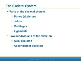
The Skeletal System • Consists of: • Bones • Joints • Cartilage • Ligaments The skeletal system consists of 206 bones.
The Skeletal System • Functions: • Bones of the lower extremities support the weight of the body. • Support and protect the soft body organs. • Enables the body to move about. • Store a number of minerals, calcium and phosphorus are most important. • Red bone marrow produces blood cells.
Classification of Bones Bones are classified as follows: • Long bones-longer than they are wide. Found in the arms and legs.
Classification of Bones • Short Bones-shaped like cubes and are found primarily in the wrists and ankles.
Classification of Bones • Flat Bones-thin, flat, curved. Form the ribs, breastbone and skull.
Classification of Bones • Irregular Bones-differently shaped, not classified as long , short, or flat. Include hip bones, vertebrae, and various bones in the skull.
Tissue and Bone Formation • Osseous tissue-another word for bone. • Osteocytes-bone cells. - secrete an intercellular matrix. -contain calcium, minerals and protein fibers.
Two Types of Bone • Compact Bone- • Dense hard bone. • Found in shafts of long bones and outer surfaces of other bones.
Two Types of Bone • Spongy Bone- • Also called cancellous bone. • Less dense • Found at the ends of long bones and in the center of other bones.
Compact Bone • Microscopically-compact bone and spongy bone look different. • Compact bone is tightly packed, density gives it strength. • Osteon or haversion system-microscopic unit of compact bone. • Each haversion system consists of mature osteocytes arranged in concentric circles around large blood vessels.
Compact Bone • Area surrounding the osteocytes is filled with protein fibers, calcium and other minerals. • Protein fibers provide elasticity. • Minerals make bone tissue hard and strong. • Compact bone consists of many haversian systems running parallel to each other, system looks like a long cylinder. • Blood vessels run laterally to the haversian system, this ensures adequate blood supply to the bone tissue.
Spongy Bone • Also called cancellous bone. • Does not have a haversian canal. • Bone tissue is arranged in plates called trabeculae. • Bony plates are separated by holes that give it a punched-out “swiss cheese” appearance. • Holes are important for: (1)decrease the weight of bone, make it lighter, and (2)contain red bone marrow.
Spongy Bone • Red bone marrow richly supplies the spongy bone with blood cells for use throughout the body. • Spongy bone is located in short, flat, and irregular bones. • Spongy bone is found in the ends of long bones.
Long Bone • Made up of an arrangement of compact and spongy tissue, which accounts for its strength. • Contains sites of growth and reshaping and structures associated with joints.
Long Bone Parts of a long bone: • Diaphysis-long shaft of the bone, composed primarily of compact bone, therefore it provides strength. • Epiphysis-enlarged ends of the long bone. Articulates or meets with a second bone at a joint. Consists of a thin layer of compact bone overlying spongy bone. Epiphysis are covered with cartilage.
Long Bone • Epiphyseal disc-band of hyaline cartilage located at each end, between the epiphysis and the diaphysis in a growing bone. This band of cartilage is the epiphyseal disc or growth plate. • Medullary cavity-hollow center of the diaphysis. The inside is lined with connective tissue called the endosteum.
Long bone • Periosteum-tough, fibrous connective tissue membrane that covers the outside of the diaphysis. • Anchored firmly to the outside of bone on all surfaces except articular cartilage. • Periosteum protects bone, serves as a point of attachment for muscle, contains blood vessels that nourish underlying bone.
Long Bone • Injury to the periosteum may have serious consequences to the health of the bone since this structure carries the blood supply. • Articular Cartilage-found on the outer surface of the epiphysis, forms a smooth shiny surface that decreases friction within a joint.
Ossification • Ossification-the formation of bone. • Occurs differently in flat and long bones. • In the fetus, flat bones in the skull consist of thin connective tissue membrane. • Ossification begins when the osteoblasts (bone forming cells), migrate to the region of the flat bones. • The osteoblasts secrete calcium and other minerals into the spaces between the membranes, thereby forming bone. • This process involves the replacement of thin membrane with bone.
Ossification of Long Bones • Ossification of long bones occurs as bone tissue replaces cartilage. • The fetal skeleton is composed largely of cartilage. • As the baby matures, osteoblasts invade the cartilage and gradually replace it with bone until all but the articular cartilage and the epiphyseal disc have been replaced by bone. • Isolated pieces of cartilage, such as the bridge of the nose and parts of the ribs remain.
Growing Bones • Two types of bone growth occurs from infancy to adulthood. (1)Longitudinally-determines the height of an individual. (2)Thicker & wider-to support the weight of the adult. • Longitudinally-bone grows at the epiphyseal disc, (also called the growth plate). • Cartilage adjacent to the epiphysis continues to multiply and grow toward the diaphysis. • Cartilage next to the diaphysis is invaded by osteoblasts and become ossified. • As long as the cartilage continues to form within the epiphyseal disc, the bone will continue to lengthen.
Growing Bones • Longitudinal bone growth ceases when the epiphyseal disc becomes ossified and fused. • Growth hormone stimulates growth at the epiphyseal disc, making the child taller. • The sex hormones estrogen and testosterone cause the epiphyseal disc to fuse, inhibiting further longitudinal growth. • The epiphyseal disc is more sensitive to estrogen, this causes girls to tend to be shorter than boys.
Growing Bones • Longitudinal growth generally ceases after puberty. • Injury to the epiphyseal disc may impair longitudinal bone growth. Eg. Injured leg shorter than uninjured leg. • Giantism- hypersecretion of growth hormone. • Dwarfism- undersecretion of growth hormone.
Growth Hormone Abnormalities Giantism Dwarfism
Growing Thicker and Wider • After longitudinal bone growth ceases, bones continue to increase in thickness and width. • Bones are continuously being reshaped. • Bone reshaping is accomplished by a combination of the actions of osteoblasts and osteoclasts. • Osteoblasts- bone forming cells. • Osteoclasts- bone destroying cells.
Growing Bones • Osteoblastic Activity: While osteoblasts build new bone, osteoclasts, found on the inner bone surface break down bone tissue, thereby hollowing out the interior of the bone. • Osteoclastic Activity is like sculpting, the bricklayer lays the bricks and the sculptor will hollow out the middle so that it is not too heavy. • Bone resorption-(not reabsorption)-process whereby osteoclasts breakdown bone matrix.
Growing Bones • Bone resorption- widens the bone, moves calcium from the bone to the blood. • Bone resorption plays a crucial role in blood calcium levels. • Weight-bearing – factor that stimulates bone growth. • Exercise and weight bearing keep calcium in the bone and increase bone mass. • Bedridden and sedentary people tend to lose bone mass causing bones to be easily broken when stressed.
Surface Markings • Surface of bones appears bumpy and irregular. • Appearance is due to numerous ridges, projections, depressions, and grooves called bone surface markings. • These bone surface markings serve as points of attachment for muscles, tendons and ligaments.
Condyle • Condyle- large rounded knob that usually articulates with another bone.
Epicondyle • Epicondyle- an enlargement near or above a condyle.
Bone Markings • Head- an enlarged and rounded end of a bone.
Bone Markings • Facet- a small flattened surface.
Bone Markings • Crest- a ridge on a bone
Bone Markings Process-a prominent projection on a bone.
Bone Markings Spine- sharp projection.
Bone Markings Tubercle (tuberosity)- a knoblike projection.
Bone Markings Trochantor- a large (tuberosity) found only on the femur.
Bone Markings Depressions/Openings Foramen- an opening through a bone, usually serves as a passageway for nerves, blood vessels, and ligaments.
Bone Markings Fossa- a depression or groove.
Bone Markings Meatus-a tunnel or tubelike passageway.
Bone Markings • Sinus-a cavity or hollow space.
Fractures Fractures- break in the bone. • Simple Fracture- a break in which the overlying skin remains intact. Local tissue damage is minimal. • Compound Fracture- a broken bone that also pierced the skin. Ends of the broken bone usually cause extensive tissue damage. • Incomplete Fracture- (greenstick)-usually occurs in children, the break is incomplete because the child’s bone is still made up of some cartilaginous material.