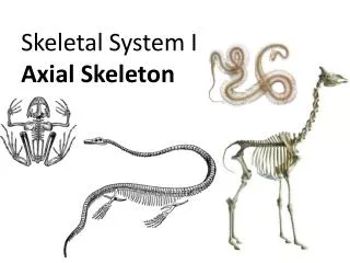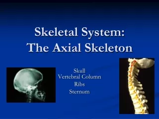
The Axial Skeletal System
The Axial Skeletal System. Divisions of the Skeletal System. Humans are born with approximately 300 bones which fuse to 206 bones as adults. There are 2 main divisions of the skeletal system: axial skeleton and appendicular skeleton. Divisions of the Skeletal System.

The Axial Skeletal System
E N D
Presentation Transcript
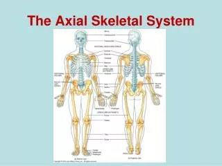
Divisions of the Skeletal System • Humans are born with approximately 300 bones which fuse to 206 bones as adults. • There are 2 main divisions of the skeletal system: axial skeleton and appendicular skeleton.
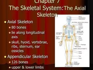
Divisions of the Skeletal System • Axial skeleton forms the vertical axis of the body. • 80 bones= skull (22), vertebral column (26), ribcage (25), auditory ossicles (6), and hyoid (1) • Appendicular skeleton forms the arms, legs and the girdles • Girdles attach the arms and legs to the axial skeleton • 126 bones= pectoral girdle (4), arms (60), legs (60) and pelvic girdle (2)
Skull • Superior end of the vertebral column • Composed of flat and irregular shaped bones • Large hollow space within the skull is called the cranial vault or cranial cavity. • Functions to: • Surround and protect the brain • Be points of attachment for the facial muscles (landmarks)
Divisions of the Skull • Cranial division consists of 8 flat bones that form a protective box around the brain. • Help to form the cranial vault (cavity) • Frontal (1): forms the anterior portion of the cranial cavity • Forms the superior orbits of the eyes and forms the forehead
Frontal Bone Landmarks • Supraorbital margin: a thickened ridge of bone found superior to the orbit of the eye. • Just deep to the eyebrow and more prominent on the lateral portion • Point for muscle attachment (PFMA) • Supraorbital foramen: a small opening found on the medial aspect of the supraorbital margin. • Can feel it best inferior to the margin • Allows blood vessels and nerves to enter the frontal bone
Frontal Bone Landmarks • Frontal sinus: A hollow space found within the frontal bone, superior and medial to the supraorbital margin. • Can only be seen with a sagittal cut • ¼ inch superior to the eyebrows • House mucus and macrophages for trapping and destroying foreign particles.
Frontal Bone Landmarks • Frontal sinus: A hollow space found within the frontal bone, superior and medial to the supraorbital margin. • Can only be seen with a sagittal cut • ¼ inch superior to the eyebrows • House mucus and macrophages for trapping and destroying foreign particles.
Parietal Bones (2) • Form the lateral walls and the superior portion of the cranium. • Landmarks: • Temporal fossa: A large, shallow depression that begins on the parietal bone and extends to the frontal bone. • PFMA
Temporal Bones (2) • Form the inferior lateral walls and a portion of the floor of the cranium. • Articulate with the mandible (lower jaw) to form the temporomandibular joint (TMJ) • Temporal bone landmarks: • Mastoid process: A large, blunt projection found posterior to the external auditory meatus. • Bump behind the ear • PFMA
Temporal Bone Landmarks cont.. • Styloid process: A thin, sharp projection found inferior and medial to the external auditory meatus. • Covered with muscle so it is more difficult to identify • PFMA • Zygomatic process: A thin, flat projection found anterior to the external auditory meatus. • PFMA • External auditory meatus: The external ear canal • Opening through which the auditory nerve runs.
Temporal Bone Landmarks cont.. • Mandibular fossa: A shallow depression found inferior and slightly anterior to the external auditory meatus • This forms an articulation with the mandible • Easy to see inferiorly if the mandible is removed
Occipital Bone (1) • Forms the posterior wall and the floor of the cranium. • The spinal cord passes through this as it exits the cranial vault. • Occipital bone landmarks: • External occipital protuberance: A prominent midline projection found on the superior surface. • Where the occipital bone turns to form the horizontal part.
Occipital Bone Landmarks cont.. • Superior nuchal line: Two curved ridges that extend laterally from the external occipital protuberance. • PFMA • Inferior nuchal line: Two curved ridges that extend laterally from the external occipital protuberance, inferior to the superior nuchal line. • PFMA • Foramen magnum: A large opening in the inferior surface of the occipital bone that allows the spinal cord to exit the cranial cavity. • Largest foramen in the body.
Occipital Bone Landmarks cont.. • Occipital condyles: Paired oval-shaped projections found lateral to the foramen magnum. • Form an articulation with the 1st bone of the spine (atlas)
Sphenoid Bone (1) • Forms the anterior floor of the cranial cavity. • Also forms a portion of the lateral walls of the cranial cavity. • Forms the posterior wall of the orbits of the eyes. • The keystone bone for the cranium because it articulates with all other cranial bones. • Shape resembles a bat with outstretched wings when viewed superiorly.
Sphenoid Bone Landmarks • Greater wing: The larger, inferior projection of the sphenoid that forms a portion of the floor and the lateral walls of the cranium. • Also forms the posterior wall of the orbits of the eyes. • Lesser wing: The smaller, superior projection of the sphenoid bone located posterior to the frontal bone.
Sphenoid Bone Landmarks cont.. • Sellaturcica: A small, saddle-like depression found between the greater and lesser wings that surrounds and protects the pituitary gland. • Pituitary is an important endocrine gland • 3 parts to the sellaturcica: • Tuberculumsellae: The anterior portion of the sellaturcica. • Closest to the lesser wing.
Sphenoid Bone Landmarks cont.. • 3 parts to the sellaturcica continued: • Hypophyseal fossa: The seat of the saddle. • Where the pituitary gland resides • Dorsum sellae: The posterior portion of the sellaturcica. • Closer to the greater wing
Ethmoid Bone (1) • The small bone located anterior to the sphenoid bone in the middle of the frontal bone. • Forms a small portion of the anterior floor of the cranium. • Also forms a small portion of the medial wall of the eye orbits. • Also forms the superior portion of the nasal septum.
Ethmoid Bone Landmarks • Cribriform plate:Paired projections found lateral to the crista galli. • Has small openings called the olfactory foramina. • Olfactory foramina: A series of small openings found within the cribriform plate that allow nerves from the olfactory epithelium to pass directly into the brain. • These nerves give us our sense of smell.
Ethmoid Bone Landmarks cont.. • Crista galli:A small triangular projection found in the center of the ethmoid bone. • Near the front of the cranial cavity. • Point of attachment for the meninges (protective coverings of the brain). • Perpendicular plate: A small vertical projection arising from the inferior surface of the ethmoid bone. • Forms the superior portion of the nasal septum. • Articulates with the vomer (facial bone).
Ethmoid Bone Landmarks cont.. • Superior and middle nasal conchae:Two thin, scroll-shaped projections found lateral to the perpendicular plate • The middle nasal conchae is inferior to the superior nasal conchae. • These increase surface area of the nasal passageways • Ethmoidal cells: Air spaces found within the lateral masses of the ethmoid bone. • Small sinuses
Sutures • Fibrous joints found between the bones of the cranium. • There are 4 major sutures: • Coronal: unites the frontal bone and both parietal bones • Sagittal: unites the two parietal bones on the superior midline of the skull • Lambdoid: unites the two parietal bones to the occipital bone. • Squamous (2): unite the parietal and temporal bones on the lateral sides of the skull
The Facial Division • A group of 14 irregular bones that serves as points of attachment for muscles of the face. • Nasal (2): form the bridge of the nose. • Rectangular shaped bones • PFMA • Maxillae (2): Form the upper jaw. • Articulate with every face bone except the lower jaw. • Form part of the floors of the orbits, lateral walls and floor of the nasal cavity, and most of the hard palate (bony roof of the mouth).
Maxillary Landmarks • Infraorbital foramen: Small openings found inferior to the orbits of the eyes. • Allows passage of blood vessels and nerves. • Palatine process: a lateral projection that forms one half of the anterior portion of the hard palate. • Typically the 2 processes unite during weeks 10-12 of embryo development. If not, cleft palate will result. This negatively impacts speech and swallowing.
Maxillary Landmarks cont… • Maxillary sinuses: a series of small spaces within the maxillae. • Empty into the nasal cavity.