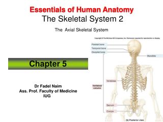
The skeletal system: the axial skeleton
860 likes | 1.19k Vues
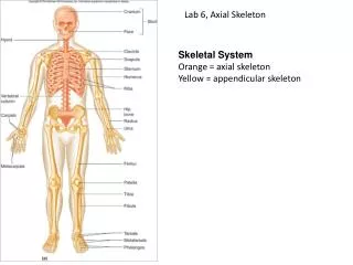
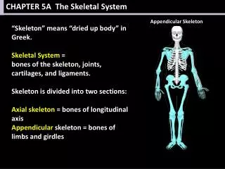
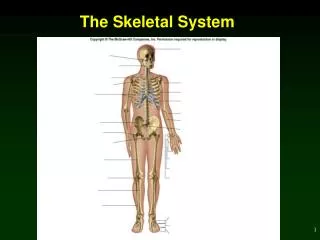
The skeletal system: the axial skeleton. Essential Question. What is the main contribution the axial skeleton makes to homeostasis?. Divisions of the Skeleton. AXIAL SKELETON Skull Cranium Face Hyoid Auditory Ossicles Vertebral Column Thorax . APPENDICULAR SKELETON: Pectoral Girdle

The skeletal system: the axial skeleton
E N D
Presentation Transcript
Essential Question • What is the main contribution the axial skeleton makes to homeostasis?
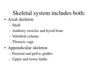
Divisions of the Skeleton • AXIAL SKELETON • Skull • Cranium • Face • Hyoid • Auditory Ossicles • Vertebral Column • Thorax • APPENDICULAR SKELETON: • Pectoral Girdle • Upper Limbs • Pelvic Girdle • Lower Limbs
Types of Bone • bones fall into 1 of 5 main types based on shape: • Long • Short • Flat • Irregular • Sesamoid
Long Bones • longer than they are wide • may be slightly curved (to absorb stress of weight at more points along the bone i.e. Straight bones would fracture more easily) • Consist of: • shaft & variable #s of ends • compact bone in diaphysis and spongy bone in epiphysis
Short Bones • somewhat cube-shaped • nearly equal in length as width • Consist of: • Spongy Bone except @ surface
Flat Bones • Composed of: • 2 nearly parallel plates of compact bone enclosing spongy bone inside • Function: • give considerable protection • place for muscle attachment
Irregular Bones • complex shapes (do not fit in other categories) • vary in amt spongy bone
Sesamoid Bones • develop w/in certain tendons where there is considerable friction, tension, & physical stress • Function: protect tendon from excessive wear & tear • vary in # person to person but everyone has 2 patella which develop in quadriceps femoris tendon
Sutural Bones • classified by location (w/in a suture: a seam between 2 cranial bones) • not everyone has them
Bone Surface Markings • 2 major types: • depressions & openings • form joints or allow passage of vessels & nerves • processes • projections or outgrowths that either help form joints or serve as attachment points for ligaments & tendons
Skull • 22 bones in 2 categories: • Cranium • 8 bones that form cranial cavity • 1 frontal bone • 2 parietal bones • 2 temporal bones • 1 occipital bone • 1 sphenoid bone • 1 ethmoid bone
Skull 2. Facial Bones 14 bones that form the face • 2 nasal bones • 2 maxillae • 1 mandible • 2 zygomatic • 2 lacrimal • 2 palantine • 2 inferior nasal conchae • 1 vomer
Skull: Cavities • Cranial Cavity • Nasal Cavity • Orbits • Paranasal Sinuses • Middle & Inner Ear Cavities
Movable Joints of the Skull • Mandible • Auditory Ossicles
Functions of Cranial Bones • protecting brain • stabilizing position of brain, vessels, & nerves through attachments to the meninges • outer surfaces provide large areas of attachment for muscles that move parts of the head & some for facial expression
Cranial Bones: Frontal Bone • forms: • forehead • upper part of eye socket • most of anterior part of cranial floor • in newborns: rt & lt which fuse shortly after birth
Parietal Bones • form greater portion of sides & roof of cranial cavity
Temporal Bones • form inferior, lateral aspects of the cranium & part of the cranial floor • its zygomaticproceess forms the lateral half of the zygomatic arch • mandibularfossa: where condylar process of mandible forms TMJ (temporal mandibular joint)
Temporal Bones • external auditory meatus: ear canal • mastoid: posterior & inferior to external auditory meatus, contains “air cells” (mastoiditis: inflammation in air cells)
Temporal Bone: Petrous Portion • base of skull between sphenoid & occipital bones • houses middle & inner ear • Carotid foramen & Jugular foramen
Temporal Bones • internal auditory meatus: passage of Cranial nerves VII (facial n.) and VIII (vestibulocochlear n.) • styloid process: point of attachment for muscles & ligaments of the tongue & neck
Occipital Bone • forms back of head & most of base of skull • foramen magnum: large hole spinal cord passes thru, • occipital condyles: articulate with 1st cervical vertebra (atlas)
Sphenoid Bone • middle base of skull • *articulates with all other cranial bones • shape resembles a bat
Sphenoid Bone • sellaturcica: (Turkish saddle) the “seat” of the saddle is the hypophysealfossa: where the pituitary gland sits • optic foramen: between body & lesser wings, cranial nerve II (optic n.) and opthlamic artery pass thru
Ethmoid Bone • “like a sieve” • midline of anterior part of cranial floor, anterior to sphenoid, posterior to nasal bones • cribiforme plate: forms roof of nasal cavity, the holes of the sieve where olfactory nerves pass from roof of nasal cavity to brain • cristagalla: triangular process which serves as pt of attachment for meninges of brain
Ethmoid Bone • perpendicular plate: forms superior portion of nasal cavity • superior & middle nasal conchae: (or turbinate) increase vascular & mucous membrane surface area in nasal cavities: aids in sense of smell, warms, filters & moistens air being inhaled. Filters because the turbinates cause air to swirl as a result inhaled particles strike & become trapped in mucus
“Sometimes when you study anatomy, you start seeing others differently.”
Facial Bones • shape of face changes dramatically during 1st 2 yrs of life: • brain & cranial bones expand • 1st set of teeth erupt • paranasal sinuses enlarge • growth of face stops ~16 years old
Facial Bones • 14 facial bones: • 2 nasal bones • 2 maxillae • 2 zygomatic bones • 1 mandible • 2 lacrimal bones • 2 palatine bones • 2 inferior nasal conchae • 1 vomer
Nasal Bones • form part of the bridge of the nose (rest is cartilage)
Maxillae • = upper jaws • *articulate with every bone in face except the mandible • form part of floor of orbits, parts of nasal cavity, & most of the hard palate (bony roof of mouth) • each one has large maxillary sinus • alveolar process is small arch that contains the alveolar sockets for upper set of teeth
Cleft Palate & Cleft Lip • 10-12 wks gestation the palatine processes of maxillae typically join • not doing so cleft palate +/- cleft lip • speech & swallowing can be affected • many ear infections • reparative surgery recommended 1st few wks of life / surgery needs to be completed by 12 – 18 mos b/4 speech: speech therapy & orthodontic care frequently necessary
Zygomatic Bones • “cheekbones”: the temporal process of the zygomatic bone articulates with the zygomatic process of the temporal bone • also part of floor of orbit
Lacrimal Bones • thin, about the size of pinky fingernail (smallest bones of face) • part of medial wall of each orbit • each contain lacrimalfossa that houses lacrimal sac: gathers tears nasal cavity