Download
1 / 49
580 likes | 1.34k Vues
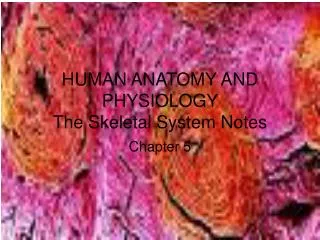
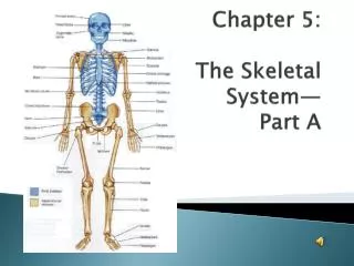
Chapter 5: The Skeletal System— Part A. The Skeletal System. Parts of the skeletal system: Bones (skeleton) Joints Cartilages Ligaments – fibrous cords that bind bones together Divided into two divisions: Axial skeleton – forms longitudinal axis (down center of body)

E N D
The Skeletal System • Parts of the skeletal system: • Bones (skeleton) • Joints • Cartilages • Ligaments – fibrous cords that bind bones together • Divided into two divisions: • Axial skeleton – forms longitudinal axis (down center of body) • Appendicular skeleton– limbs & girdles (attachment areas of limbs to axial skeleton)
Skeletal Bones 206 total bones in body 80 126
Functions of Bones • Support of the body • Protection of soft organs • Movement due to skeletal muscles attached by tendons • Storage of minerals (Ca & P) and fats • Blood cell formation = hematopoeisis
The skullprotects the brain Bones protect soft organs…
Bones of the Human Body • The adult skeleton has 206 bones • Two basic types of bone tissue: • Compact bone (dense) • Homogeneous • Spongy bone • Small needle-like pieces of bone • Many open spaces Figure 5.2b
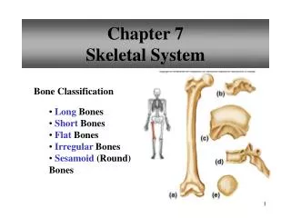
Classification of Bones by Shape • Four types of bone: Long Short Flat Irregular
Classification of Bones on the Basis of Shape Figure 5.1 Hoban
Classification of Bones • Long Bones • Typically longer than wide • Have a shaft with heads at both ends • Containmostly compact bone • Examples: Femur, humerus • Includes all bones of limbs except wrist & ankle
Femur Note the thinner shaft area and the wider heads at both ends
Classification of Bones • Short Bones • Generally cube-shape • Contain mostly spongy bone • Examples: Carpals (wrist), tarsals (ankle) • Sesamoidbonesfound in tendons-- ex. patella
Classification of Bones • Flat Bones • Thin and flattened • Usually curved • Two thin layers of compact bone around a layer of spongy bone • Examples: Skull, ribs, sternum
Classification of Bones • Irregular Bones • Irregular shape • Do not fit into other categories • Example: Vertebrae and hip
Gross Anatomy of a Long Bone • Diaphysis • Shaft • Composed of compact bone • Epiphysis • Ends of the bone • Composed mostly of spongy bone covered with thin layer of compact bone • Covered with articular (glassy hyaline) cartilage Figure 5.2a
Structures of a Long Bone • Periosteum • Outside covering of the diaphysis • Fibrous connective tissue membrane • Sharpey’s Fibers • Secure periosteum to underlying bone • Arteries • Supply bone cells with nutrients Figure 5.2c
Structures of a Long Bone • Articular cartilage • Covers the external surface of the epiphyses • Made of hyaline cartilage • Decreases friction at joint surfaces Figure 5.2a
Anatomy of a Long Bone • Epiphyseal plate • Flat plate of hyaline cartilage seen in young, growing bone • Epiphyseal line • Remnant of the epiphyseal plate • Seen in adult bones
Structures of a Long Bone • Medullary Cavity • Cavity of the shaft • Contains yellow marrow (mostly fat) in adults • Contains red marrow (for blood cell formation) in infants Figure 5.2a
Bone Markings • Surface features of bones • Sites of attachments for muscles, tendons, and ligaments • Passages for nerves and blood vessels • Categories of bone markings • Projections and processes – grow out from the bone surface • Depressions or cavities – indentations
Microscopic Anatomy of Bone • Osteon(Haversian System) • A unit of bone containing central canal and matrix rings • Haversian canal, osteocytes (mature bone cell), lacunae, lamellae • Central (Haversian) canal (run lengthwise) • Opening in the center of an osteon • Carries blood vessels and nerves • Perforating (Volkman’s) canal • Canal perpendicular to the central canal • Carries blood vessels and nerves
Microscopic Anatomy of Bone • Lacunae • Cavities containing bone cells (osteocytes) • Arranged in concentric rings • Lamellae • Rings around the central canal • Sites of lacunae Detail of Figure 5.3 Hoban
Canaliculi • Tiny canals • Radiating outward from central canal to lacunae • Form a transport system connecting all bone cells to a nutrient supply • Calcium salts give hardness, while collagen fibers give flexibility
Changes in the Human Skeleton • In embryos, the skeleton is primarily hyaline cartilage • During development, much of this cartilage is replaced by bone • Cartilage remains in isolated areas • Bridge of the nose • Parts of ribs • Joints Hoban
Changes in the Human Skeleton • Flat bones form on fibrous membranes • Most bones develop using hyaline structures as models = ossification • Ossification • Cartilage covered with bone matrix from osteoblasts • Inner cartilage digested leaving medullary cavity • Happens by birth Hoban
Bone Growth (Ossification) • Epiphyseal plates allow for lengthwise growth of long bone during childhood • New cartilage is continuously formed • Older cartilage becomes ossified • Cartilage is broken down • Enclosed cartilage is digested away, opening up a medullary cavity • Bone replaces cartilage through the action of osteoblasts
Bone Growth (Ossification) • Bones are remodeled and lengthened until growth stops • Bones are remodeled in response to two factors • Blood calcium levels • Pull of gravity and muscles on the skeleton • Bones grow in width (called appositional growth)
Bone startingto replacecartilage Bone collar Hyalinecartilagemodel In an embryo (a) Long Bone Formation and Growth Figure 5.4a, step 1
Hyalinecartilage New center ofbone growth Medullarycavity Bone startingto replacecartilage Bloodvessels Growthin bonelength Bone collar Hyalinecartilagemodel In an embryo In a fetus (a) Long Bone Formation and Growth Figure 5.4a, step 2
Articularcartilage Hyalinecartilage Spongybone New center ofbone growth New boneforming Epiphysealplatecartilage Growthin bonewidth Medullarycavity Bone startingto replacecartilage Bloodvessels Growthin bonelength New boneforming Bone collar Hyalinecartilagemodel Epiphysealplate cartilage In an embryo In a fetus In a child (a) Long Bone Formation and Growth Figure 5.4a, step 3
Long Bone Formation and Growth Figure 5.4b
Types of Bone Cells • Osteocytes—mature bone cells • Osteoblasts—bone-forming cells • Osteoclasts—bone-destroyingcells • Break down bone matrix for remodeling and release of calcium in response to parathyroid hormone • Bone remodeling is performed by both osteoblasts and osteoclasts
Long Bone Formation & Growth • Controlled by hormones • Growth hormone • Sex hormones (puberty) • Ends during adolescence • Epiphyseal plates converted to bone
Bone Remodeling • Process by both osteoblasts and osteoclasts • Cells regenerate in response to blood Ca levels & pull of gravity & muscles on skeleton • Osteoclasts • Stimulated by parathyroid (in throat) hormone to break down bone & release Ca to blood • Hypercalcium • Increased blood Ca causes calcium to be deposited in bone • Stress on bone • Causes bones to thicken & the projection to increase size where muscles are attached
Bone Formation Issue: Rickets Softened bones bow due to decreasedcalcium or decreased vitamin D (needed to absorb calcium) Results from severe malnutrition in early life Treated by increasing Calcium, Phosphorus, and Vitamin D in diet, as well as exposure to sun.