Chapter 7 Skeletal System
Chapter 7 Skeletal System. Bone Classification. Long Bones Short Bones Flat Bones Irregular Bones Sesamoid (Round) Bones. Skeletal System. Functions Support Protection internal organs Movement facilitation Bones act as the levers Articulations as the fulcrums

Chapter 7 Skeletal System
E N D
Presentation Transcript
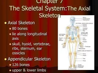
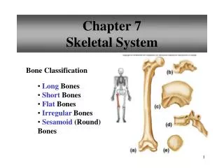
Chapter 7Skeletal System Bone Classification • Long Bones • Short Bones • Flat Bones • Irregular Bones • Sesamoid (Round) • Bones
Skeletal System • Functions • Support • Protection • internal organs • Movement facilitation • Bones act as the levers • Articulations as the fulcrums • Muscles provide the force
Skeletal System • Functions • Mineral Storage • calcium and phosphorous • Storage of Blood Cell Producing Cells • red bone marrow • Storage of Energy • lipids stored in yellow bone marrow are important source of chemical energy
Bone Function • Support, Movement & Protection • gives shape to head, etc. • supports body’s weight • protects lungs, etc. • bones and muscles interact • when limbs or body parts move • Inorganic Salt Storage • calcium • phosphate • magnesium • sodium • potassium • Blood Cell Formation • hematopoiesis • red marrow
Cartilage, Osseous and Dense CT Osseous Tissue (4 cell types) Osteoprogenitor Found throughout the bone Have mitotic potential May differentiate into osteoblast Cells and Histology of Bone
Cells and Histology of Bone • Osseous Tissue (4 cell types) • Osteoblast • No mitotic potential • Found on the surface of the bone • Secrete mineral salts and organic components for bone formation
Cells and Histology of Bone • Osseous Tissue (4 cell types) • Osteocytes • No mitotic potential • Found within the bone • Maintain daily cellular activities of bone tissue • Osteoclast • Found on the surface of bone • Function in the re-absorption of bone
Parts of a Long Bone • Epiphysis • distal end • proximal end • Articular Cartilage • Covers each epiphysis • Hyaline cartilage • Diaphysis • Shaft • Metaphysis • Area between diaphysis and epiphysis in mature bone
Periosteum Contains nerves, blood vessels, osteo cells Attachment point for ligaments and tendons Medullary cavity contains yellow marrow Endosteum lines the medullary cavity Parts of a Long Bone
SpongyBone Irregular lattice work of bone-trabeculae Spaces filled with red marrow produces RBCs Parts of a Long Bone
Compact Bone Tightly packed tissue Parts Concentric Lamellae Concentric circles of tightly packed tissue Volkmann’s Canals Penetrate compact bone Allow blood vessels and nerves into medullary cavity and other Haversian Canals Parts of a Long Bone
Compact Bone Haversian Canals Run longitudinally through the compact bone Concentric lamellae surround the canals Lacunae Open spaces between the concentric lamellae that contain osteocytes Look like “little lakes” Parts of a Long Bone
Compact Bone Canaliculi Tiny canals that radiate away from the lacunae Osteon (Haversian System) Central canal, surrounding lamellae, lacunae, osteocytes and canaliculi Parts of a Long Bone
Bone Development • Intramembranous Ossification • bones originate within sheetlike layers of connective tissues • broad, flat bones • skull bones (except mandible) • intramembranous bones • Endochondral Ossification • bones begin as hyaline cartilage • form models for future bones • most bones of the skeleton • endochondral bones
Endochondral Ossification • hyaline cartilage model • primary ossification center • secondary ossification centers • epiphyseal plate • osteoblasts vs. osteoclasts
Hyaline cartilage model “bone” created by chondroblast Actual ossification of this “bone” starts 6-7 weeks after conception Endochondral Ossification
Endochondral Ossification • Interstitial and Oppositional Growth • Chondrocytes burst and trigger calcification • Capillaries and bone cells invade dying cartilage • Bone starts to calcify
Primary Ossification Center Blood vessels in mid-region produce ossification center Osteoblast form spongy bone in the area of calcified cartilage, this causes the center to enlarge Spongy bone is destroyed by osteoclast-forms medullary cavity Endochondral Ossification
Secondary Ossification Centers When blood vessels enter the epiphysis Spongy bone created here will stay Epiphyseal plate Layer of hyaline cartilage between the 2 areas of growth Stays until you reach maturity Endochondral Ossification
Remnants of hyaline cartilage on the epiphysis becomes articulating cartilage Endochondral Ossification
Growth at the Epiphyseal Plate • First layer of cells • closest to the end of epiphysis • resting cells • anchors epiphyseal plate to epiphysis • Second layer of cells • many rows of young cells • undergoing mitosis
Growth at the Epiphyseal Plate • Third layer of cells • older cells • left behind when new cells appear • cells enlarging and becoming calcified • Fourth layer of cells • thin • dead cells • calcified extracellular matrix
Homeostasis of Bone Tissue • Bone Re-absorption – action of osteoclasts and parathyroid hormone • Bone Deposition – action of osteoblasts and calcitonin
Factors Affecting Bone Development, Growth, and Repair • Deficiency of Vitamin A – retards bone development • Deficiency of Vitamin C – results in fragile bones • Deficiency of Vitamin D – rickets, osteomalacia • Insufficient Growth Hormone – dwarfism • Excessive Growth Hormone – gigantism, acromegaly • Insufficient Thyroid Hormone – delays bone growth • Sex Hormones – promote bone formation; stimulate ossification of epiphyseal plates • Physical Stress – stimulates bone growth
Superficial Bone Anatomy • Process • bony prominence “bump” • Condyle • rounded or knuckle like process • Tubercle • small process • Tuberosity • large process (tibial tuberosity) • Trochanter • huge process (located on the proximal end of the femur)
Superficial Bone Anatomy • Crest • narrow ridge of bone • Spine • sharp slender process (holes and/or depressions) • Fissure • narrow slit through which blood vessels or nerves pass • Foramen • opening through which blood vessels and nerves pass
Superficial Bone Anatomy • Meatus • tubelike passageway running within a bone • Sulcus or groove • furrow that accommodates a soft structure such as blood vessels, nerves or tendons • Fossa • depression in or on the surface of a bone
Life-Span Changes • decrease in height at about age 30 • calcium levels fall • bones become brittle • osteoclasts outnumber osteoblasts • spongy bone weakens before compact bone • bone loss rapid in menopausal women • hip fractures common • vertebral compression fractures common
Impacted one fragment is firmly driven into the other Displaced anatomical alignment is NOT preserved Non-displaced anatomical alignment is preserved Stress partial fracture resulting in bones inability to withstand forces About 25% involve distal end of the fibula Clinical Application: Fractures
Clinical Application Types of Fractures • green stick • fissured • comminuted • transverse • oblique • spiral
Clinical Application: Fractures • Pathologic • caused by weakening of the bone due to a disease • Pott’s • fracture of the distal end of the fibula with serious injury to the distal tibial articulation • Severe eversion sprain may lead to this • Colle’s • fracture at the distal end of the radius in which the distal end is displaced posteriorly • Occurs frequently when you try to stop yourself from falling
Fracture breaks blood vessels found in the Haversian system Blood clot forms at the site of the break within 6 to 8 hours (fracture hematoma) Fracture hematoma serves as the focus for cellular invasion Clinical Application: Fracture Repair
Callus forms New bone tissue developed around the fracture Site of osteoblast activity Clinical Application: Fracture Repair
Remodeling Dead bone is absorbed by osteoclast Compact bone replaces spongy bone in the fractured area Clinical Application: Fracture Repair
Osteoporosis Age related disease characterized by decreased bone mass an increased chance of fractures Decreased levels of estrogen (sex hormones that stimulate osteoblast) Clinical Application: Disorders
Clinical Application: Disorders • Osteoporosis • White women more than men and people of color • Other factors linked to osteoporosis • Body build • short people have less bone mass • Weight • thin people have less adipose, which stores estrogen • Smokers- • low estrogen levels • Calcium deficiency and mal-absorption • Vitamin D deficiency • Certain drugs- • alcohol, cortisone, etc. • Premature menopause
Clinical Application: Disorders • Osteoporosis Prevention • Exercise • muscle action stimulates blood flow to bone tissue • Estrogen pills- • for post menopause women • Calcium supplements/ vitamins • Prescription anabolic steroids- • increase hormone levels • Sodium Fluoride stimulates oteoblast
Clinical Application: Disorders • Osteogenic Sarcoma • Malignant bone tumor that affects osteoblast • People between ages of 10-25 • Left untreated it will metasticize and lead to death • Treatment includes chemotherapy following amputation of affected area
Clinical Application: Disorders • Rickets • Vitamin D deficiency in children • Body can’t transport Ca+2 and P-3 from GI tract to blood • Osteoblasts in diaphysis don’t calcify causing bones to stay soft • Weight of body causes the legs to bow • Cure and prevention • Dietary- add vitamin D, calcium and phosphorus in large amounts • Exposing the skin to ultraviolet rays of light
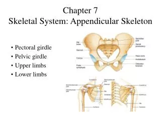
Skeletal Organization • Axial Skeleton • head • neck • trunk • Appendicular Skeleton • upper limbs • lower limbs • pectoral girdle • pelvic girdle
Skeletal Organization • Hyoid Bone • Supports tongue • Attachment point for muscles that move tongue
Cranium • Frontal(1) • Forehead • Roof of nasal cavity • Roofs of optical orbits • Frontal sinuses • Supraorbital foramen • Blood vessels and nerve • Coronal suture • Develops in two parts • Completely fused by age 5 or 6
Cranium • Parietal(2) • Side walls of cranium • Roof of cranium • sagittal suture • Fuses two parietal bones together • Coronal suture • Fuses parietal bones to frontal bone
Cranium • Occipital (1) • back of skull • base of cranium • foramen magnum • Inferior part of brain stem connects with spinal cord • occipital condyles • Either side of foramen magnum • lambdoid suture • Fuses parietal bones with occipital bone
Cranium • Temporal (2) • side walls of cranium • floor of cranium • floors and sides of orbits • squamous suture • Fuses parietal with temporal • external acoustic meatus • Leads to inner ear • mandibular fossa • Where mandible meets temporal bone • mastoid process • Neck muscle attachment • styloid process • Tongue and pharynx attachment • zygomatic process
Cranium • Sphenoid (1) • base of cranium • sides of skull • floors and sides of orbits • sella turcica • Indentation along midline within cranial cavity • Contains pituitary gland • sphenoidal sinuses
Cranium • Ethmoid(1) • roof and walls of nasal cavity • floor of cranium • wall of orbits • cribriform plates • Join two parts of ethmoid on either side of nasal cavity • perpendicular plate • Forms most of nasal septum • superior and middle nasal conchae • Support mucous membranes of nasal cavity • ethmoidal sinuses • crista galli • Projects upward into cranial cavity • Attaches brain membranes
Facial Skeleton • Maxillary (2) • upper jaw • anterior roof of mouth • floors of orbits • sides of nasal cavity • floors of nasal cavity • alveolar processes • maxillary sinuses • palatine process