Skeletal System Functions in Human Bones
Learn about the functions and classifications of the skeletal system, including the structure of long bones and the roles of osteoblasts and osteoclasts. Explore bone development processes and factors affecting bone growth. Discover fracture types and repair stages. Dive into the world of bone health.

Skeletal System Functions in Human Bones
E N D
Presentation Transcript
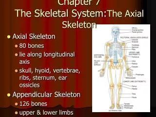
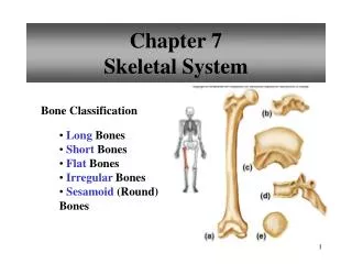
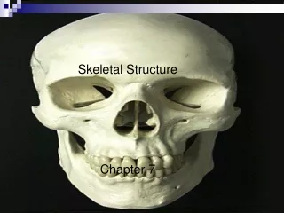
Chapter 7 Skeletal System
Functions of Skeletal System:1. Support 2. Protection3. Produces blood cells4. Stores inorganic materials
Classification – bones classified by shape: • 1. Long • 2. Short • 3. Flat • 4. Irregular • 5. Sesamoid
Structure of Long Bone Epiphysis- expanded portion at end, forms joint. Epiphyseal disk (line)-growth plate Diaphysis- shaft of bone Medullary cavity-hollow chamber in center Spongy bone- cancellous bone, found mainly in epiphysis Compact bone-tightly packed tissue Periosteum-tough, vascular covering Endosteum- membrane of bone forming cells Articular cartilage-layer of hyaline cartilage on epiphysis of bone
Structure of a Long Bone • Red marrow- formation of red blood cells, white blood cells and platelets. • Found in flat bones and epiphysis • Yellow marrow- stores fats, no blood cell production. • Found in diaphysis
Compact vs. Spongy Bone • Perforating(Volkmann’s) canal- Connects central canals • Trabeculae-bony plates of spongy bone • Canaliculi- canals btwn osteocytes • Osteocyte-Bone cells • Lacunae- space in bone containing osteocytes
Bone Structure Animation • structure of bone animation
Osteoblast vs. Osteoclast • Osteoblast- bone forming cells which deposit Calcium and Phosphorous into the dense collagen framework. • Osteoclast- Large multinucleated cells formed from fusing white blood cells. • Secrete an acid which breaks down the bony matrix • Both work together over a person’s lifetime to remodel bone and allow for resorption on minerals.
Osteoblast vs osteoclast • osteoblast osteoclast animation
Bone Development – 2 Types: 1. Intramembranous ossification (changing cartilage to bone) (ex. - skull) • layers of undifferentiated connective tissue appear at sites of future bone • connective tissue cells differentiate into osteoblasts (cells that build bone) around blood vessels • osteoblasts become osteocytes (mature bone cells) when completely surrounded by a bony matrix • Connective tissue on surface forms the periosteum
2. Endochondral ossification (remainder of skeleton) • Hyaline cartilage models form at sites of future bone • Cartilage cells degenerate over time • osteoblasts form bone over cartilage • osteoblasts become osteocytes when completely surrounded by a bony matrix
Ossification Animation • Ossification animation
Bone Development • In a long bone, hyaline cartilage is replaced by bony tissue in the center of the diaphysis first (primary ossification center) • Bone develops from this point towards the end of the bone • Secondary ossification centers appear in the epiphysis last.
Growth of Bones • Epiphyseal disk – area of growth; separates epiphysis from diaphysis; 4 layers: 1. Resting cells – closest to end; do not add length but act as anchor 2. Reproducing cells - as they reproduce, they lengthen the bone 1.
Growth of Bones 3. Mature cells – growing and accumulating Ca 4. Degenerating cells & osteoblasts depositing bony tissue
Epiphyseal Plate Video • http://www.youtube.com/watch?v=RTx-Z0a9wvM
Epiphyseal disk • Bones continue to lengthen until epi. disks are ossified (approx. 21 yrs. of age) • Bones thicken through- out life as compact bone is added under peri- osteum • Hyaline cartilage on ends remains
Bone Growth Video • http://app.discoveryeducation.com/player/view/assetGuid/39B53919-B11C-4D2C-A92C-E030DF639351
Epiphyseal disk 1. Radial growth plate→ 2. Fracture – distal end of radius → Digital growth plate (in fingers) (2 ½ yr. old)→
Fractures • Greenstick- incomplete break in convex surface • Fissured- incomplete longitudinal break • Comminuted-complete, fragments bone • Transverse- complete break at right angle • Oblique- complete at angle other than right angle • Spiral- complete, caused by twisting of bone
Fracture Repair • 4 stages • Blood escapes from blood vessels and from hematoma. • Spongy bone forms near blood vessels, fibrocartilage forms in distant regions (within days to weeks) • Bony callus forms from cartilaginous callus similar to bone formation • Osteoclasts remove excess bone to restore to original
Bone Fracture Video • https://www.youtube.com/watch?v=MNkI6Of2PRs
Factors Affecting Bone Growth(Hormones) Deficiency Effect Pituitary growth Pituitary dwarfism hormone (excess – pituitary (stimulates cell division gigantism/acro- in the epi. disks) megaly) Thyroid hormone premature disk (stimulates cartilage ossification-growth replacement in disk) stunt ed
Acromegaly Yao Defen is 34 yrs. old & The world’s tallest female at 7ft. 8 in!!
Factors Affecting Bone Growth(Hormones & Exercise) Deficiency Effect Sex hormones stimulate ossifi- (cause growth of cation of disks; long bones; estrogen stop bone length stronger than androgen) Physical stress bone thins & weak- (stimulates bone width; ens; atrophy hypertrophy)
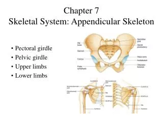
2 divisions of skeleton: • Axial – skull, vertebrae, sternum, ribs (thorax), hyoid bone • Appendicular – pectoral & pelvic girdles, upper & lower limbs Human Skeleton – composed of 206 bones (extra – sutural)
Surface Markings on Bone • Surface of bones have various structural features for specific functions • Depressions & Openings: Foramen Meatus Sinus
Foramen Opening for b.v., nerves, ligaments (ex.-foramen magnum)
Meatus Tubelike passageway (ex.-external auditory meatus)
Sinus Cavity within a bone (ex.-frontal sinus)
Surface Markings on Bone • Processes that Form Joints: Condyle Head Facet
Condyle Rounded process for articulation w/another bone (ex. – mandibular condyle)
Head An enlargement at the end of a bone (ex.-head of femur)
Facet Small, nearly flat surface where 2 bones articulate (ex.-vertebral facet)
Surface Markings on Bone • Processes that Connective Tissue Attaches To: Tuberosity Spine Trochanter Crest Process
Tuberosity Knoblike process (ex.-ischial tuberosity)
Spine Thornlike ridge or projection (ex.-spine of scapula)
Trochanter Large process or projection (ex.-trochanter of femur)
Crest Ridgelike projection (ex.-iliac crest)
Process Prominent projection on a bone (ex.-spinous process)