Download
1 / 38
380 likes | 408 Vues
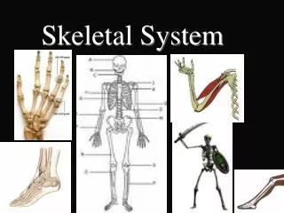
Learn about the structure and functions of the pelvic girdle bones, hip bones, femur, tibia, fibula, foot bones, and joints in the skeletal system. Understand the differences between male and female pelves. Discover the significance of arches in the foot.

E N D
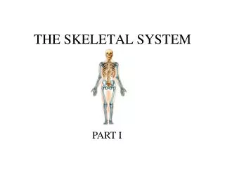
Bones of the Pelvic Girdle • Pelvic Girdle - Formed by two coxal bones, or ossa coxae, commonly called the hip bones. • Bony Pelvis – Hip bones + coccyx + sacrum
Bones of the Pelvic Girdle • Large and heavy bones, and they are attached to the axial skeleton. • The sockets, which receive the thigh bones, are deep and heavily reinforced by ligaments that attach the limbs firmly to the girdle. • Functions of the Girdle: • Bearing weight is the most important function. • Protect the reproductive organs, urinary bladder, and part of he large intestine.
Structure of the Hip Bones • Each hip bone is formed by the fusion of three bones: • Ilium • Ischium • Pubis
Ilium • Ilium – Large, flaring bone that forms most of the hip bone. • Connects posteriorly with the sacrum at the sacroiliac joint. • When you put your hands on your hips, they are resting over the winglike portion of the ilia. • Iliac Crest – The upper edge of the winglike portion of the ilium.
Ischium • Ischium – “Sitdown bone”; Forms the the most inferior part of the coxal bone. • Pubis – Most anterior part of a coxal bone.
Acetabulum • Acetabulum – Deep socket that receives the head of the thigh bone. • Formed by the fusion of the ilium, ischium, and pubis.
Regions of the Bony Pelvis • False Pelvis – Superior to the true pelvis; The area medial to the flaring portions of the ilia. • True Pelvis - Surrounded by bone; Lies inferior to the flaring parts of the ilia and the pelvic brim. • Dimensions of the true pelvis of a woman are very important because they must be large enough to allow the infant’s head to pass during childbirth.
Differences Between a Male and Female Pelvis • The pelvis of a female tends to be: • Inlet is larger and more circular. • As a whole is shallower, and the bones are lighter and thinner. • Ilia flare more laterally. • Sacrum is shorter and less curved. • Ischial spines are shorter and farther apart; thus the outlet is larger. • Pubic arch is more rounded because the angle of the pubic arch is greater.
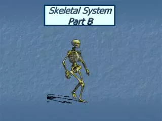
Bones of the Lower Limbs • The lower limbs carry our total body weight when we are erect. • Hence, it is not surprising that the bones of the lower limbs are much thicker and stronger than the comparable bones of the upper limb. • The 3 segments of the lower limbs: • Thigh • Leg • Foot
Thigh • Femur – Thigh bone. • Only bone in the thigh. • The heaviest, strongest bone in the body. • It slants medially as it runs downward to join with the leg bones. • This brings the knees in line with the body’s center of gravity. • The medial course of the femur is more noticeable in females because of the wider female pelvis.
Structure of the Femur Bone • Its proximal end has a: • Ball-like head • The head of the femur articulates with the acetabulum of the hip bone in a deep, secure socket. • A neck • Common site of fractures, especially in old age.
The Distal Femur • Anteriorly on the distal femur is the smooth patellar surface, which forms a joint with the patella (kneecap).
Leg • Two bones form the skeleton of the leg: • Tibia • Fibula • The tibia and fibula are connected along their length by an interosseous membrane.
Tibia and Fibula • Tibia – Shinbone; Larger and more medial. • At the proximal end, the tibia articulates with the distal end of the femur to form the knee joint. • Fibula – Lies alongside the tibia; Thin and sticklike. • Forms joints with the tibia both proximally and distally. • Has no part in forming the knee joint. • The distal end of the fibula forms the outer part of the ankle.
Foot • The foot is composed of the: • Tarsals • Metatarsals • Phalanges • Two important functions of the foot: • Supports our body weight • Serves as a lever that allows us to propel our bodies forward when we walk or run.
Foot: Tarsals • Tarsus – The posterior half of the foot. • Composed of 7 tarsal bones. • Body weight is mostly carried by the two largest tarsals: • Calcaneus - Heelbone • Talus – Tarsal that lies between the tibia and the calcaneus.
Foot: Metatarsals and Phalanges • The sole of the foot: • Composed of 5 metatarsals. • The toes of the foot: • Composed of 14 phalanges. • Like the fingers of the hand, each toe has three phalanges, except the great toe which has two.
Arches of the Foot • The bones in the foot are arranged to form three strong arches: • Two longitudinal (medial and lateral) • One Transverse • Ligaments (bind the foot bones together) and tendons: • Help to hold the bones firmly in the arched position but still allow a certain amount of give or springiness.
Joints • Every bone in the body (except the hyoid bone of the neck) forms a joint with at least one other bone. • Joints (Articulations) – Sites where two or more bones meet.
Functions of Joints • Joints have two functions: • Hold the bones together securely • Give the rigid skeleton mobility • Joints are classified in two ways: • Functionally (focuses on the amount of movement allowed by the joint) • Structurally
Functional Classification of Joints 1. Synarthroses: Immoveable joints;Allows no movement. - Example: Bones in the skull 2. Amphiarthroses– Slightly moveable joints; Allow a small amount of restricted movement. - Example: Vertebrae and the joints between the two bones of the lower leg. 3. Diarthroses - Freely Moveable; Permit movement in one or more directions - Example: Shoulder, neck, and knee
Structural Classification of Joints • Structurally, there are three types of joints: • Fibrous Joints • Cartilagenous Joints • Synovial Joints
Fibrous Joints • Fibrous Joints – The bones are united by fibrous tissue; As a general rule, these are immoveable. • Bones are bound tightly together by CT fibers, allowing essentially no movement. • Example: Sutures of the skull.
Cartilagenous Joints • Cartilagenous Joints – Bone ends are connected by cartilage. • Slightly Moveable Examples: • Pubic symphysis of the pelvis • Intervertebral joints of the spinal column (connected by discs of fibrocartilage) • Immoveable Examples: • The Epiphyseal plates of growing long bones • Joints between the first ribs and the sternum. • Most cartilagenous joints are slightly moveable.
Synovial Joints • Synovial Joints – Joints in which the articulating bone ends are separated by a joint cavity containing synovial fluid.
All synovial joints have four distinguishing characteristics: 1. Articular cartilage • Covers the ends of the bones forming the joint. 2. Fibrous articular capsule • Joint surfaces are enclosed by a sleeve or capsule of fibrous CT • The capsule is lined with a smooth synovial membrane (the reason these joints are called synovial joints).
All synovial joints have four distinguishing characteristics (continued): 3. Joint cavity • The articular capsule encloses a cavity, called the joint cavity, which contains lubricating synovial fluid. 4. Reinforcing ligaments • The fibrous capsule is usually reinforced with ligaments.
Bursae and Tendon Sheaths • Not strictly part of synovial joints, but they are often found closely associated with them. • Bursae – Flattened fibrous sacs lined with synovial membrane and containing a thin film of synovial fluid. • Common where ligaments, muscles, skin, tendons, or bones rub together. • Tendon Sheath – An elongated bursae that wraps completely around a tendon subjected to friction. • Like a bun around a hot dog.
Types of Synovial Joints: Based on Shape • Plane • Hinge • Pivot • Condyloid • Saddle • Ball-and-socket
Plane Joint • Plane Joint – The articular surfaces are essentially flat, and only short slipping or gliding movements are allowed. • Movements are nonaxial (does not involve rotation around any axis). • Examples: Intercarpal joints of the wrist.
Hinge Joint • Hinge Joint – The cylindrical end of one bone fits into a trough-shaped surface on another bone. • Angular movement is allowed in just one plane, like a mechanical hinge. • Classified as uniaxial (they allow movement around one axis only). • Examples: elbow joint, ankle joint, and the joints between the phalanges of the fingers.
Pivot Joint • Pivot Joint – The rounded end of one bone fits into a sleeve or ring of bone. • Because the rotating bone can turn only around its long axis, pivot joints are also uniaxial joints. • Examples: • Proximal radioulnar joint • Joint between the atlas and the dens of the axis
Condyloid Joint • Condyloid Joint – The egg-shaped articular surface of one bone fits into an oval concavity in another. • Both of these articular surfaces are oval. • Allow the moving joint to travel (1) from side to side and (2) back and forth. • But the bone cannot rotate around its long axis. • Movement occurs around two axes, hence these joints are biaxial.
Saddle Joint • Saddle Joint – Each articular surface has both convex and concave areas, like a saddle. • These biaxial joints allow essentially the same movements as condyloid joints. • Example: Carpometacarpal joints in the thumbs
Ball-and-Socket Joint • Ball-and-Socket Joint - the spherical head of one bone fits into a round socket in another. • These multiaxial joints allow movement in all axes, including rotation and are the most freely moving synovial joints. • Examples: Shoulder and hip
Dislocations • A dislocation happens when a bone is forced out of its normal position in the joint cavity. • Reduction – The process of returning the bone to its proper position. • Should be done only by a physician. • Attempts by an untrained person to “snap the bone back into its socket” are usually more harmful than helpful.
Sprains • Sprains – The ligaments or tendons reinforcing a joint are damaged by excessive stretching, or they are torn away from the bone. • Since tendons and ligaments get poor blood supply, sprains heal slowly and are extremely painful.