Download
1 / 70
760 likes | 1.13k Vues
Discover the skeletal system, its parts, bones, joints, cartilages, ligaments, and classifications based on shapes. Learn about bone growth, bone cells, fractures, and the healing process. Understand the axial skeleton, including details about the skull. Dive into bone formations, types of bone cells, and the stages of bone fracture healing.

E N D
Chapter 5The Skeletal System Day 1 Notes:
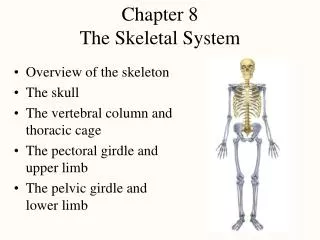
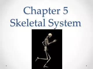
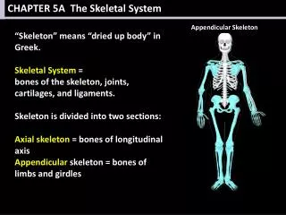
The Skeletal System • Parts of the skeletal system • Bones (skeleton) • Joints • Cartilages • Ligaments (bone to bone)(tendon=bone to muscle) • Divided into two divisions • Axial skeleton: bones of the skull, vertebral column, and rib cage • Appendicular skeleton: bones of the upper and lower limbs, shoulder and hip
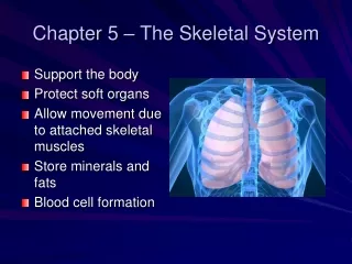
Functions of Bones • Support of the body • Protection of soft organs • Movement due to attached skeletal muscles • Storage of minerals and fats • Blood cell formation
Bones of the Human Body • The adult skeleton has 206 bones • Two basic types of bone tissue • Compact bone • Homogeneous • Spongy bone • Small needle-like pieces of bone • Many open spaces
Classification of Bones • 1. Long bones • Typically longer than wide • Have a shaft with heads at both ends • Contain mostly compact bone • Examples: Femur, humerus
Classification of Bones • 2. Short bones • Generally cube-shape • Contain mostly spongy bone • Examples: Carpals, tarsals
Classification of Bones • 3. Flat bones • Thin and flattened • Usually curved • Thin layers of compact bone around a layer of spongy bone • Examples: Skull, ribs, sternum
Classification of Bones • 4. Irregular bones • Irregular shape • Do not fit into other bone classification categories • Example: Vertebrae and hip
Gross Anatomy of a Long Bone • Diaphysis • Shaft (middle) • Composed of compact bone • Epiphysis • Ends of the bone • Composed mostly of spongy bone Figure 5.2a
Structures of a Long Bone • 1. Periosteum • Outside covering of the diaphysis • Fibrous connective tissue membrane • 2. Sharpey’s fibers • Secure periosteum to underlying bone • 3. Arteries • Supply bone cells with nutrients Figure 5.2c
Structures of a Long Bone • 4. Articular cartilage • Covers the external surface of the epiphyses • Made of hyaline cartilage • Decreases friction at joint surfaces Figure 5.2a
Structures of a Long Bone • 5. Medullary cavity • Cavity of the shaft • Contains yellow marrow (mostly fat) in adults • Contains red marrow (for blood cell formation) in infants Figure 5.2a
Bone Markings • Surface features of bones • Sites of attachments for muscles, tendons, and ligaments • Passages for nerves and blood vessels • Categories of bone markings • Projections and processes – grow out from the bone surface • Depressions or cavities – indentations
Changes in the Human Skeleton • In embryos, the skeleton is primarily hyaline cartilage • During development, much of this cartilage is replaced by bone • Cartilage remains in isolated areas • Bridge of the nose • Parts of ribs • Joints
Bone Growth • Epiphyseal plates allow for growth of long bone during childhood • New cartilage is continuously formed • Older cartilage becomes ossified • Cartilage is broken down • Bone replaces cartilage
Bone Growth • Bones are remodeled and lengthened until growth stops • Bones change shape somewhat • Bones grow in width
Long Bone Formation and Growth Figure 5.4a
Types of Bone Cells • Osteocytes • Mature bone cells • Osteoblasts • Bone-forming cells • Osteoclasts • Bone-destroying cells • Break down bone matrix for remodeling and release of calcium
STOP: What’s Next? • -Bone Fractures • -How Bones Heal • -Axial Skeleton • -The Skull-
Bone Fractures • A break in a bone • Types of bone fractures • Closed (simple) fracture – break that does not penetrate the skin • Open (compound) fracture – broken bone penetrates through the skin • Bone fractures are treated by reduction and immobilization • Realignment of the bone
Common Types of Fractures Table 5.2
Repair of Bone Fractures • Hematoma (blood-filled swelling) is formed • Break is splinted by fibrocartilage to form a callus • Fibrocartilage callus is replaced by a bony callus • Bony callus is remodeled to form a permanent patch
Stages in the Healing of a Bone Fracture Figure 5.5
The Axial Skeleton • Divided into three parts • Skull • Vertebral column • Bony thorax
The Skull • Two sets of bones • Cranium • Facial bones • Bones are joined by sutures • Only the mandible is attached by a freely movable joint
The Skull Figure 5.7
Bones of the Skull Figure 5.11
Human Skull, Superior View Figure 5.8
Human Skull, Inferior View Figure 5.9
Paranasal Sinuses • Hollow portions of bones surrounding the nasal cavity Figure 5.10
Paranasal Sinuses • Functions of paranasal sinuses • Lighten the skull • Give resonance and amplification to voice Figure 5.10
The Hyoid Bone • The only bone that does not articulate (move) with another bone • Serves as a moveable base for the tongue Figure 5.12
The Fetal Skull • The fetal skull is large compared to the infants total body length Figure 5.13
The Fetal Skull • Fontanelles – fibrous membranes connecting the cranial bones • Allow the brain to grow • Convert to bone within 24 months after birth Figure 5.13
Stop: What’s Next? The Upper half of the body
The Vertebral Column • Vertebrae separated by intervertebral discs • The spine has a normal curvature • Each vertebrae is given a name according to its location Figure 5.14
Structure of a Typical Vertebrae Figure 5.16
The Bony Thorax • Forms a cage to protect major organs Figure 5.19a
The Bony Thorax • Made-up of three parts • Sternum • Ribs • Thoracic vertebrae Figure 5.19a
The Appendicular Skeleton • Limbs (appendages) • Pectoral girdle • Pelvic girdle
The Appendicular Skeleton Figure 5.6c
The Pectoral (Shoulder) Girdle • Composed of two bones • Clavicle – collarbone • Scapula – shoulder blade • These bones allow the upper limb to have exceptionally free movement
Bones of the Shoulder Girdle Figure 5.20a, b
Bones of the Upper Limb • The arm is formed by a single bone • Humerus Figure 5.21a, b
Bones of the Upper Limb • The forearm has two bones • Ulna • Radius Figure 5.21c
Bones of the Upper Limb • The hand • Carpals – wrist • Metacarpals – palm • Phalanges – fingers Figure 5.22
Bones of the Pelvic Girdle • Hip bones • Composed of three pair of fused bones • Ilium • Ischium • Pubic bone • The total weight of the upper body rests on the pelvis • Protects several organs • Reproductive organs • Urinary bladder • Part of the large intestine
The Pelvis Figure 5.23a
Gender Differences of the Pelvis Figure 5.23c