Download
1 / 20
200 likes | 493 Vues
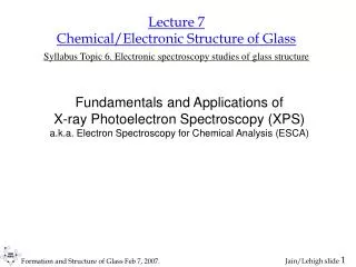
Lecture 7 Chemical/Electronic Structure of Glass Syllabus Topic 6. Electronic spectroscopy studies of glass structure. Fundamentals and Applications of X-ray Photoelectron Spectroscopy (XPS) a.k.a. Electron Spectroscopy for Chemical Analysis (ESCA). Bibliography.

E N D
Lecture 7Chemical/Electronic Structure of GlassSyllabus Topic 6. Electronic spectroscopy studies of glass structure Fundamentals and Applications of X-ray Photoelectron Spectroscopy (XPS) a.k.a. Electron Spectroscopy for Chemical Analysis (ESCA)
Bibliography • D. Briggs and M.P. Seah, Practical surface analysis, vol 1. Auger and XPS. Wiley, 1990. • M.A. Sherwood in Surface imaging and visualization, A.T. Hubbard, ed. Ch 63. CRC Press. • D.A. Skoog, F.J. Holler, T.A. Nieman, Principles of Instrumental Analysis, Brooks/Cole. Ch. 21. • NIST X-ray Photoelectron Spectroscopy Database: http://srdata.nist.gov/xps/ • http://www.emsl.pnl.gov/new/emsl2002/tutorials/engelhard_xps.pdf • www.courses.vcu.edu/PHYS661/pdf/08TechSpectroscopy041.ppt
Outline • Introduction – chemical structure, electron spectroscopy XPS as a tool for composition analysis Surface limitation of XPS Chemical structure • Spectrometer • Video tour of Scienta instrument • http://rm1.cc.lehigh.edu:8080/asxgen/dept/IMI/VirtualGlassCourse/Video/XPS_miller150.wmv Charging issues - special to glass
Different Aspects of the Static Structure • Chemical structure • Nature of bonding (covalency, ionicity, basicity, etc...) between different kinds of atoms. • Charge distribution. • Physical structurePhysical arrangement of atoms with respect to each other (RM-O, DsM-O2, CN, etc..) • Short range order. • Medium range structure. • Vibrational structure • Bond strength • Local vibrations of the mobile atom. • Vibrations of network structural units of small and medium size.
There are different manifestations of the same overall glass structure The various aspects are strongly interdependent, yet we examine one aspect at a time due to the limitations of the techniques and our own vision!
Electron Spectroscopy Techniques • Photon in, Electron out • Photoemission Spectroscopy (PES)X-ray Photoemission Spectroscopy (XPS - 200 to 2,000 eV source)Ultraviolet Photoemission Spectroscopy (UPS - 10 to 50 eV source) • Auger Electron Spectroscopy (AES) • Electron in, Electron out (inelastic) • Auger Electron Spectroscopy (AES) • Electron Energy Loss Spectroscopy (EELS)
XPS: basic process • Developed in the mid 1960s by K. Siegbahn et al. • Awarded the Nobel Prize for Physics in 1981
Auger spectroscopy Auger electron: KL1L2,3 ESCA => XPS + Auger Auger (vs. XPS): Independent of input energy, Sensitivity for low Z atoms, High spatial resolution if e are used as excitation beam, Minimal matrix effects, Difficult quantitative analysis.
XPS: What do we detect? K.E. = hn – BE; if BE is w.r.t. vacuum level Often for solids BE is defined w.r.t. Fermi level. The measured KE should be corrected for contact potential (i.e. Dwork function of sample and spectrometer), and any surface charging of an insulating sample. Then K.E. = hn - BE - f(sample) - f(spectro.) - S e: discrete peak, e’: background that decreases with increasing k.e. Also a step at each peak. XPS info is from 10-100 A, depending on k.e.
Relative intensity of XPS signal from different shells • Calculated cross sections (1.5 keV photon energy) give probabilities for observing electrons from various energy levels. • The intensity of XPS signal decreases with increasing shell, the valence band being the weakest.
Binding energy of electrons in various elements BE for a particular electron shell is characteristic of the element => Identify the element from its BE. However, watch out for interference as the BE of different shell electrons of different elements may overlap!
Core levels and Auger Survey spectrum Identification of almost all elements in one experiments Valence band Survey spectrum of an in situ fractured sodium diborate (Na2O-2B2O3) glass
Core level peak attributes: • Height • Area • Asymmetry • Position • Spin-orbit split (for higher than s-levels) W 4f core level spectrum of W metal exposed to air. Proctor and Sherwood, Anal. Chem. (1982).
What can we learn from core level spectrum? • Compared to valence electrons, the core levels are little affected by the bonding • Core level BE is characteristic of the element, but it is chemically shifted (0 to 10 eV) by interaction with valence electrons and surrounding (mostly nearest neighbor) atoms. • Symmetric for insulator but asymmetric for metals from excitation of conduction electrons. • (p3/2, p1/2), (d5/2, d3/2), (f7/2, f5/2).. levels show two peaks from spin-orbit coupling. The ratio of respective peaks are: 2/1; 3/2; 4/3 => Core level XPS gives qualitative and quantitative chemical structure around particular element (excluding H and He). • The area under a core level peak is concentration of the element => Get composition using reference sensitivity factors.
1 2 3 4 5 Variation of surface composition
Effect of corrosion on surface composition: leaching vs. uniform dissolution • After washing • Na • Ca • Al • Si/O • Rim is richer in alumina.
XPS sees only the surface region (XPS info is from 10-100 A, depending on k.e.)
Glass composition: surface vs. bulkHigh resolution X-ray photoelectron spectroscopy Preferential loss of alkali and alkaline earth oxide from the surface.
Electron detector Take-off angle = 5 x-ray beam Laser light beam x-ray beam Laser light beam Electron detector Take-off angle = 90 specimen specimen Surface sensitivity => Angle Resolved XPS Obtain depth profile of the chemical info! (XPS info is from 10-100 A, depending on k.e.)
Composition as f(depth, ambient) As50Se50 film on Si, irrad. in air for ~12 h