Download
1 / 76
760 likes | 914 Vues
RESPIRATORY SYSTEM Department of Anatomic Pathology Faculty of Medicine Brawijaya University. Tingkat Kemampuan 2

E N D
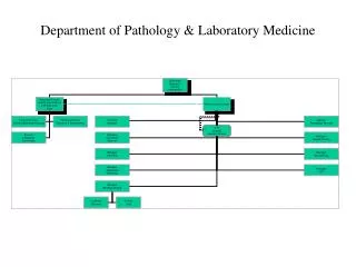
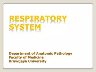
RESPIRATORY SYSTEMDepartment of Anatomic PathologyFaculty of MedicineBrawijaya University
Tingkat Kemampuan 2 Lulusandoktermampumembuat diagnosis klinikterhadappenyakittersebutdanmenentukanrujukanbagipasienkepadaspesialis yang relevan. Lulusandokterjugaharusmampumenindaklanjutisesudahnya. Tingkat Kemampuan 3 3A. Lulusandoktermampumemberikanpenangananterhadappenyakittersebutpadakeadaan yang bukankeadaangawatdaruratsertamampumemberikanterapipendahuluandemimenyelamatkannyawaataumencegahkeparahandanataukecacatanpadapasien. Lulusandokterharusdapatmenentukanrujukanyang paling tepatbagipenangananlebihlanjutuntukpasiendanmampumenanganiperawatanselanjutnya.
3B. Lulusandoktermampumemberikanpenanganangawatdaruratpadapenyakittersebutdanmampumemberikanterpaipendahuluandemimenyelamatkannyawaataumencegahkeparahandanataukecacatanpadapasien. Lulusandokterharusdapatmenentukanrujukan yang paling tepatbagipenangananlebihlanjutuntukpasiendanmampumenanganiperawatanselanjutnya. Tingkat Kemampuan 4 Lulusandoktermampumenanganipenyakittersebutsecaramandiridantuntas. Merekaharusmampumenegakkan diagnosis pasienberdasarkanpemeriksaanfisikdanpemeriksaanpenunjang (misalnya: pemeriksaanlaboratoriumsederhanaatauX-ray) yang tepatgunadantidakberlebihan.
KOMPETENSI Diagnosis Rujuk Diagnosis 1st Treatment (Non Emergency) rujuk Diagnosis 1st treatment (Emergency) Rujuk Diagnosis Appropriate Treatment
DISEASES of RESPIRATORY SYSTEM • ATELECTASIS • COPD (EMPHYSEMA) • PULMONARY TUBERCULOSIS • LUNG CANCER • Definition • Pathophysiology • Macros • Micros • X-Ray • Clinical Appearance • Management
What is it? # Atelectasis (at-uh-LEK-tuh-sis) is a condition in which one or more areas of lungs collapse or don't inflate properly. # Incomplete expansion of the lung (neonatal atelectasis) or collapse of previously inflated lung (acquired atelectasis), producing areas of relatively airless pulmonary parenchyma # Reversible
Possible Underlying Disease Lung diseases such as COPD, asthma, cystic fibrosis, tuberculosis, and whooping cough also increase your risk for a collapsed lung
Cause of Atelectase • Acquired atelectasis • resorption ( obstruction) atelectasis • compression atelectasis • contraction atelectasis
Resorption atelectasis * Complete obstruction of an airway resorption of the oxygen trapped in the dependent alveoli lung volume diminished mediastinum shifts toward the atelectatic lung * Cause : excessive secretions (e.g., mucus plugs) or exudates within smaller bronchi most often found in bronchial asthma, chronic bronchitis, bronchiectasis, postoperative states, aspiration of foreign bodies, bronchial neoplasms (rarely)
Compression atelectasis * Cause : the pleural cavity is partially or completely filled by fluid exudate, tumor, blood, or air (pneumothorax) mediastinum shifts away from the affected lung Contraction atelectasis * Cause : local or generalized fibrotic changes in the lung or pleura prevent full expansion
Resorption Compression Contraction
Clinical Feature cough, but not prominent chest pain breathing difficulty low oxygen saturation pleural effusion (transudate type) cyanosis (late sign) increased heart rate
Management • Based on the predetermined cause • Non Pharmacological non Invasive • chest physiotherapy : including postural drainage, chest wall percussion and vibration, and a forced expiration technique (called huffing) • Non Pharmacological invasive • Suctioning • Bronchoscopy • Pharmacological • Antibiotic if there is evident of infection • Nebulized bronchodilators and humidity may help liquefy secretions and promote their easy removal
Learning Resources http://emedicine.medscape.com/article/296468-overview
COPD (chronic obstructive pulmonary disease ) * Emphysema (acinar level) * Chronic bronchitis (bronchial level) - Commonly caused by cigarette smoking - The fourth leading cause of morbidity and mortality in USA
Emphysema : Condition of the lung characterized by irreversible enlargement of the airspaces distal to the terminal bronchiole, accompanied by destruction of their walls without obvious fibrosis
Types of Emphysema (according to its anatomic distribution within the lobule) Four major types : (1) Centriacinar(centrilobular) (2) Panacinar(panlobular) (3) Paraseptal(distal acinar) (4) Irregular * Centriacinar and panacinar cause clinically significant airflow obstruction * Centriacinar emphysema : > 95% of cases
Centriacinar Emphysema The central or proximal parts of the acini, formed by respiratory bronchioles, are affected, whereas distal alveoli are spared - > severe in upper lobes, particularly apical segments - Walls of the emphysematous spaces : black pigment (++) - Inflammation around bronchi and bronchioles (+) - Severe distal acinus may also be involved differentiation from panacinar emphysema : difficult - >> in heavy smokers, often in association with chronic bronchitis
Panacinar Emphysema Theacini are uniformly enlarged from the level of the respiratory bronchiole to the terminal blind alveoli - The prefix "pan" refers to the entire acinus but not to the entire lung - Tends to occur more commonly in the lower zones and in the anterior margins of the lung, and usually most severe at the bases. - Associated with α1-antitrypsin (α1-AT) deficiency
Paraseptal Emphysema Theproximal portion of the acinus is normal, and the distal part is predominantly involved - More striking adjacent to the pleura, along the lobular connective tissue septa, and at the margins of the lobules - Occurs adjacent to areas of fibrosis, scarring, or atelectasis and is usually more severe in the upper half of the lungs - Multiple, continuous, enlarged airspaces from < 0.5 cm to > 2.0 cm in diameter, sometimes forming cystlike structures
Irregular Emphysema (Airspace Enlargement with Fibrosis) The acinus is irregularly involved, almost invariably associated with scarring - In most instances, are asymptomatic and clinically insignificant
Other forms of emphysema Compensatory Hyperinflation /Emphysema - Dilation of alveoli but not destruction of septal walls in response to loss of lung substance elsewhere Obstructive Overinflation - Caused by subtotal obstruction by a tumor or foreign object
Bullous Emphysema - Large subpleural blebs or bullae ( Ø >1 cm ) - Occur in any form of emphysema - Occur near the apex, sometimes in relation to old tuberculous scarring. - Sometimes : rupture pneumothorax Interstitial Emphysema * Entrance of air into connective tissue stroma of the lung, mediastinum, or subcutaneous tissue * Caused by : - alveolar tears in pulmonary emphysema - fractured rib that punctures the lung
Possible Cause/Risk Factor Smoking Prolonged infection
Pathogenesis # The protease-antiprotease imbalance hypothesis - Genetic deficiency of the antiprotease α1- antitrypsin (normally present in serum, tissue fluids, and macrophages, a major inhibitor of proteases particularly elastase) - Activated neutrophils (in smokers) ROS inactivation of α1-antitrypsin elastase activity ↑ tissue damage pulmonary emphysema
# Smoking * Neutrophils and macrophages accumulate in alveoli - Chemoattractant effects of nicotine - Effects of ROS (free radicals) contained in smoke activated neutrophils /macrophages proteases (elastase, etc) release tissue damage * Oxidant-antioxidant imbalance - Tobacco smoke : ROS/free radicals (++) deplete antioxidant mechanisms tissue damage * Activated neutrophils ROS inactivation of α1-antitrypsin
Clinical course : Appear if > ⅓ of pulmonary parenchyma is damaged - Dyspnea - Cough - Wheezing confused with asthma - Weight loss - Barrel-chest - Expiratory airflow limitation best measured through spirometry (the key to diagnosis)
PULMONARY TUBERCULOSIS • Caused by : Mycobacterium Tuberculosis • Tissue reaction : - Granulomatous reaction (tubercle): - Hard tubercle - Soft tubercle • Mode of transmission : 1. airborne dropled 2. food 3. skin 4. feces
Primary TBC * Develops in a previously unexposed, and therefore unsensitized person * Usually occur in children & infant * Sometimes occur in previously unexposed or profoundly immunosuppressedadults
Pathogenesis : * Bacilli lower part of the upper lobe/upper part of the lower lobe (close to the pleura) sensitization Ghon focus(1- 1.5 cm area of gray white inflammatory consolidation, caseous necrosis +) regional nodes +, >>caseate (+) Ghon complex(GF+ regional nodes) * In the first few weeks lymphatic/ hematogenous spread may occur. * ± 95% of cases cell-mediated immunity Ghon complex fibrosis calcification
MACROS : The initial lesion : - Ø<2 cm, 1-2 cm of the apical pleura. - Sharply circumscribed, firm, gray-white to yellow - Central caseation, peripheral fibrosis - Fibrous encapsulation fibrocalcific scars MICROS : -Coalescent tubercles with central caseation -Heal with fibrosis : spontaneously / tx (+) -Progress and extend along several different pathways